Equine Clinical Case Challenge: More than Just a Corneal Lesion?

A 17-year-old Quarterhorse gelding was presented for a corneal lesion of approximately five months duration. The owner reported that the lesion started as a faint gray to white haziness then became pinker in appearance. Treatment with topical steroids produced no apparent change in the lesion. Cytology was obtained via corneal scraping but was nondiagnostic and the horse was referred to a specialist in the American College of Veterinary Ophthalmologists for further evaluation.
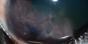
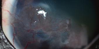
Ocular examination revealed a superficial vascularized lesion affecting a large segment of the ventral cornea (Figures 1 and 2). The surface of the lesion had an irregular or pebbled texture. The lesion did not appear to affect the surrounding conjunctiva and the remainder of the ocular examination was unremarkable. The eye was comfortable and visual.
What are your differential diagnoses and what are your next steps?
The primary differential diagnosis for a corneal lesion with a papillary appearance is squamous cell carcinoma (SCC). SCC is more commonly associated with conjunctival lesions but horses may also present with isolated corneal involvement. Less likely differential diagnoses would include eosinophilic keratitis or other nonulcerative keratitides, although the appearance of the corneal lesion in this horse makes SCC by far the most likely diagnosis.
Cytology may help in ruling out eosinophilic keratitis but is frequently nondiagnostic for SCC. A corneal biopsy can provide a definitive diagnosis but is not a necessary preoperative step in a lesion with this appearance.
What is your treatment plan? What should be discussed with the owner?
Ocular surface SCC in horses rarely metastasize but may be locally destructive and invasive. Early, aggressive treatment is key to a good outcome (i.e. retention of a visual globe without tumor recurrence). Surgery alone may be appropriate for very small lesions but recurrence rates decrease significantly with the use of an adjunctive modality such as cryotherapy, radiofrequency hyperthermia, CO2 laser ablation, beta irradiation (strontium brachytherapy), or intraoperative application of mitomycin C. Regardless of the modality chosen, however, the owner should be counseled that recurrence is still a possibility and that some horses will develop bilateral disease. Post-treatment corneal scarring may also affect the horse’s vision.
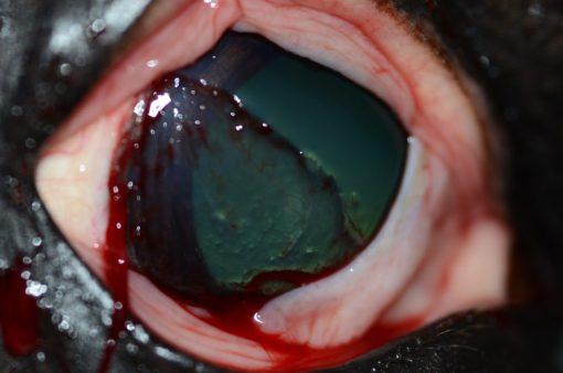
A standing superficial keratectomy was performed in this horse, incorporating 1-2 mm of clear cornea surrounding the lesion (Figure 3). Conjunctiva adjacent to the lesion was also resected despite its apparently normal appearance. The wound bed was treated with two cycles of spray cryotherapy using liquid nitrogen. A subpalpebral lavage system was placed. Postoperative treatment consisted of topical ofloxacin, autologous serum, and atropine, along with oral flunixin meglumine.
The resected corneal and conjunctival tissue was submitted for histopathology. The corneal lesion was consistent with SCC in situ – the tumor involved only the epithelium and had not breached the epithelial basement membrane – while the conjunctival tissue was considered normal. In situ SCC may represent an earlier stage of disease and may carry a better prognosis for nonrecurrence but outcomes have not been compared between horses with in situ disease and tumors that invade the corneal stroma.
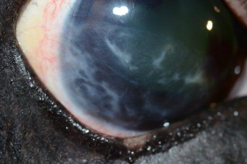
The horse was rechecked two weeks after surgery. The cornea was fully epithelialized with extensive fibrosis but no evidence of recurrent neoplasia (Figure 4). The subpalpebral lavage system was removed. To further limit the chances of recurrence, topical mitomycin C was prescribed, to be used three times a day for one week followed by a week without treatment. This cycle was to be repeated a total of three times. Mitomycin C is preferred over 5-fluorouracil as a topical chemotherapeutic as it is better tolerated by the patient and less toxic should accidental exposure to children or other animals occur. COX-2 inhibiting NSAIDs such as piroxicam may also be useful in preventing recurrence but were not selected for this horse.
Because SCC has been linked to ultraviolet light exposure, the owner was also instructed to use a UV-blocking flymask during turnout. Finally, a regular ocular examination by the family veterinarian was also recommended. As of one year postoperatively, this horse was reportedly doing well with only minor scarring at the surgical site.