Severe Keratoconjunctivitis Sicca Accompanied by Ipsilateral Xeromycteria
Background:
A 12-year-old male castrated Labrador Retriever presented to the internal medicine service with a two-month history of chronic right-sided nasal discharge refractory to antibiotic therapy. The referring veterinarian suggested computed tomography (CT) and rhinoscopy. The owners also reported mucoid to mucopurulent discharge from the right eye (OD) and occasional blepharospasm. The dog was otherwise healthy. The admitting internist noted corneal changes OD and requested an ophthalmology consult. What’s Your Diagnosis?
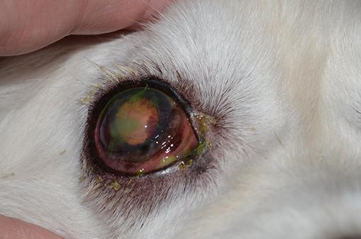
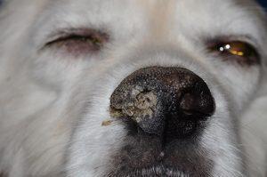
On evaluation, the dog displayed mild to moderate blepharospasm OD with thick mucopurulent discharge adherent to the lids. There were an axial superficial ulcer and moderate peripheral neovascularization along with thickened hyperemic conjunctiva OD (Figure 1). The left eye (OS) appeared normal. Schirmer tear test values were 0 mm/60 sec OD and 15 mm/45 sec OS. Intraocular pressure was normal in both eyes and both eyes are visual. The right half of the nasal planum was dry and the right nostril was occluded with dried debris (Figure 2).
What are your differential diagnoses and what are your next diagnostic steps?
This dog has unilateral severe keratoconjunctivitis sicca (KCS) accompanied by ipsilateral xeromycteria (dry nose). Taken in combination, these findings are pathognomonic for neurogenic KCS.
The most common form of KCS in dogs is bilateral and has presumed immune-mediated pathogenesis. Neurogenic KCS, however, is usually unilateral and occurs due to a failure of parasympathetic innervation to the lacrimal glands. In health, the lacrimal glands are supplied by the parasympathetic division of the facial nerve (CN VII). The parasympathetic fibers travel with the motor fibers of CN VII through the petrous temporal bone and split off at the geniculate ganglion. They travel from there in company with sympathetic fibers via the pterygoid canal and pterygopalatine fossa to the lacrimal glands, eventually joining with the zygomaticotemporal nerve, which arises from the maxillary division of the trigeminal nerve (CN V).
The parasympathetic division of CN VII also supplies the glands of the nasal mucosa, leading to the xeromycteria that accompanies neurogenic KCS. Dogs with neurogenic KCS are frequently misdiagnosed with nasal disease due to the dramatic buildup of dried debris that can occur with this condition.
Neurogenic KCS can occur due to infection, neoplasia, or trauma along any portion of the nerve. Performing a thorough neurologic examination may help to localize the lesion (it is important to note that the use of topical medications such as proparacaine or tropicamide during an ocular examination may affect the results of a subsequent neurologic examination). If ipsilateral facial nerve paralysis is also noted, then the lesion is proximal to the geniculate ganglion. More distal lesions may also produce a loss of facial anesthesia or Horner’s syndrome depending on the involvement of CN V or the sympathetic nerves. Most commonly, however, neurogenic KCS is an isolated finding and is idiopathic in origin.
In the case of this dog, a neurologic examination yielded no additional findings and a complete physical examination was likewise unremarkable. As there is a weak association between neurogenic KCS and thyroid disease, a complete blood count, chemistry panel, and thyroid panel were ordered. Results were within normal limits for all parameters. Magnetic resonance imaging (MRI) of the brain and cranial nerves was discussed with the owners but was declined due to cost.
What is your treatment plan? What should be discussed with the owner?
Neurogenic KCS does not respond to immunomodulatory lacrimal stimulant medications like cyclosporine or tacrolimus, although they are used to improve comfort in some dogs. Lubricants and cholinergic drugs are the mainstays of therapy.
Pilocarpine, a cholinergic agonist, is used both topically and orally to stimulate tear production. Topical pilocarpine can cause significant irritation and its efficacy has been questioned, although many ophthalmologists find it useful in the treatment of neurogenic KCS.
Pilocarpine is more commonly administered orally. Dosing in this manner increases the risk of side effects but has the benefit of also improving xeromycteria. Dogs require a small dose of the drug and the commercially available pilocarpine tablets intended for people cannot be used safely. Pilocarpine eye drops, on the other hand, can be administered in food to provide small, easily adjusted doses. Administering the drug in food rather than directly into the mouth is recommended to avoid an inadvertent overdose. Both 1% and 2% formulations are available; the usual starting dose is one drop of 2% pilocarpine per 10 kg of body weight given twice a day. For very small dogs 0.25% pilocarpine can be obtained from compounding pharmacies.
Systemic side effects are a concern with cholinergic agonists and may include excess salivation, vomiting, diarrhea, bradycardia, weakness, or wobbliness. Owners should be counseled to monitor for signs and to guard carefully against overdose or double-dosing. Pilocarpine should not be used in animals with cardiac disease or chronic gastrointestinal issues.
The Labrador described above was treated with oral pilocarpine along with topical tobramycin three times a day and lubricating drops as often as possible. He was rechecked one week after the commencement of therapy. At that time the tear production OD had normalized, the xeromycteria had resolved, and the ulcer had healed. The pilocarpine was continued, as most dogs seem to require long-term therapy for neurogenic KCS. The owners were advised to watch for changes in neurologic status or overall health and to return for a recheck in 6 months. At the time of recheck the dog was tolerating pilocarpine well and had experienced pilocarpine well and had experienced no recurrence of the KCS or xerostomia.