Before surgery, Otis would get winded when he was playing, but since the procedure he's been asymptomatic. We're not sure if he'll live a normal life, but he's not limited by what he went through.
Mending a Broken Heart
Domestic short-haired kitten survives innovative procedure for rare congenital heart defect
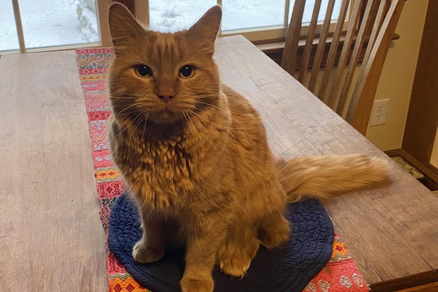
He arrived at Cummings School of Veterinary Medicine’s Henry and Lois Foster Hospital for Small Animals with an uncommon congenital heart defect. Otis, an eight-month-old orange domestic short-haired kitten, came from a Vermont-based organization called From Feral to Fur-Ever Rescue. John Rush, D.V.M., DACVIM (Cardiology), DACVECC, and Emily Karlin, D.V.M., DACVIM (Cardiology),V08 (she/her), took the lead on his treatment.
Rush, a professor, and Karlin, an assistant clinical professor, are members of Cummings School’s Department of Clinical Sciences and Foster Hospital for Small Animal’s Cardiology Service.
“The defect Otis had, Cor Triatriatum Sinister, is fairly rare,” Karlin explains. “It means three atria, but there aren’t three. A membrane inside the left atrium divides it into two compartments.”
The membrane does not have a big hole for blood to pass through, which creates a high-pressure chamber. Meanwhile, the blood has trouble getting to the low-pressure chamber on its way into the left ventricle. “This tiny narrowing tends to cause issues for kittens very early in life,” says Karlin. “It causes severe pulmonary edema (congestion) and pulmonary hypertension because the high pressure is trying to pass through the little hole.”
Unfortunately, because the membrane is difficult to access, medical options to address the issue are scarce, according to Karlin. “For most right heart diseases, you go in through the jugular vein, and it takes you directly to the heart,” she explains. “But the left atrium is not easily accessible from the outside of the body, so surgical options are quite challenging. And cats are small and usually young when they are diagnosed, so there are many logistical issues.”
After deciding against attempting a hybrid procedure through the left atrial appendage due to its complexity and potential complications, the team decided to try a different approach that had been successful in dogs with mitral valve disease.
This involves accessing the mitral valve from the apex of the left ventricle, through the ventricular myocardium (the heart’s wall). The hybrid procedure calls for a small thoracotomy, in which access is gained to the pleural space of the chest. A surgeon, Dr. Robert McCarthy, V83—retired associate clinical professor, recipient of the 2022 Cummings School of Veterinary Medicine Outstanding Alumni Award, and a 2021 inductee into the Faculty Hall of Fame—joined the collaborative effort to care for Otis, along with an anesthesia team led by Dr. Rebecca Reader, V13. “When you can see the bottom of the heart, you go in with the catheter through the left ventricle to get to the mitral valve,” Karlin says.
“Then you could put something across the little opening in the left atrium to stretch the narrowed area. You stretch it with a balloon in the end. So first you put a guide wire across it, and then you stretch it with a balloon.”
Karlin and her surgical team did not know if the procedure would be successful, but if left untreated, Otis would succumb to the disease within his first year or two of life. The surgical team shared its treatment plan with the shelter. According to Karlin, a representative from the shelter said, ‘If you think there’s a chance it could help him, or if there’s any chance that you could learn something to help future animals, please do it.’ Through fundraising efforts, From Feral to Fur-Ever Rescue collected enough money from generous donors to pay for the surgery.
Rush and Karlin estimated that Otis had about a 50 percent chance of surviving, and he did very well through the procedure. “We stretched open the membranes, so there’s still an abnormal membrane there, but it’s open wider than it was, which left less obstruction to blood flow,” Karlin explains.
After pulling through the surgery, Otis was still without a home. Enamored by him, Karlin adopted Otis and brought him home, where he joined two dogs and her husband, Mike. “Before surgery, Otis would get winded when he was playing, but since the procedure he’s been asymptomatic. We’re not sure if he’ll live a normal life, but he’s not limited by what he went through.”
In reviewing the procedure Karlin believes that this option is something that the surgical team would strongly consider if presented with a similar case. “It’s not something that we see regularly, but I think it went well enough that we would try it again for other kittens in the future. And we’re very grateful to the rescue for securing the funds to give us a chance to improve Otis’s life.”
Serving dogs, cats, and exotic pets, Henry and Lois Foster Hospital for Small Animals is one of only 15% of companion animal hospitals in North America to achieve accreditation from the American Animal Hospital Association. Designated as a Certified Level 1 facility with emergency services available 24 hours a day, Foster Hospital works with referring veterinarians and is one the nation’s top veterinary trauma centers.
Department:
Foster Hospital for Small Animals