First person: Katelin Murphy
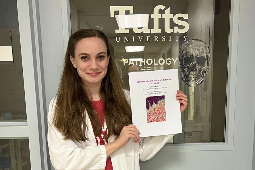
Katelin Murphy, HT (ASCP), a histotechnology professional at Cummings School of Veterinary Medicine, recently won the 2023 Art of the Stain Contest, sponsored by the National Society for Histotechnology in celebration of Histology Professionals Day. She holds a degree in histotechnology from SUNY Cobleskill and recently shared the following about her profession and the contest she won by creating art from slides.
Histotechnology professionals (also known as histotechs) prepare surgical samples for microscopic evaluation by a pathologist. This involves the processing, cutting, and staining of tissue samples so that doctors can see the cells under the microscope. This process is essential to the diagnosis of cancerous or atypical cells within the sample.
Veterinary histology comes with its own challenges, since there is such a wide variety of species from which we see samples. The histotechs and pathologists on our campus work hard to ensure your pet receives the best possible care. If your pet has had surgery at Cummings School, the tissue was most likely handled by our histology team.
The field of histology is a wonderful blend of both art and science. Each year, the National Society for Histotechnology hosts the “Art of the Stain Contest” to celebrate all the hardworking histotechs and the beautiful slides that they create.
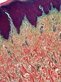
Histology professionals nationwide submit images of their slides to the contest, and the public votes for their favorite. I am honored that an image I submitted was selected as the winner of this year’s contest.
This image is the maxilla skin from a dog stained with Movat's Pentachrome technique. This procedure stains multiple structures within the tissue, such as collagen (red), mucins (blue), red blood cells (yellow), and muscle (orange). This is particularly useful when studying the heart, blood vessels, and other vascular diseases within the body.
For more information about Katie, her work in the Veterinary Diagnostic Laboratory, and her beautiful slides, visit @histoqueenofhearts on Instagram.