Ferret with a History of Vomiting

Background:
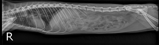
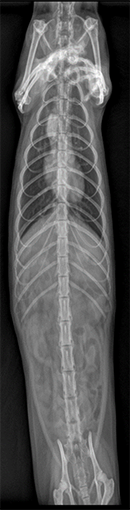
A one-year-old 0.62-kg (1.4-lb) spayed female ferret, Beyoncé, was referred to Foster Hospital for Small Animals for evaluation of a five-day history of vomiting. The ferret had been examined by the referring veterinarian 3 days prior when radiographs of the thorax and abdomen were obtained (Figures 1 and 2), and amoxicillin-clavulanic acid was prescribed to treat gastroenteritis. On initial physical examination, the ferret was alert, responsive and normothermic; however, it was intermittently retching during handling and slight pulmonary crackles were evident on caudal thoracic auscultation bilaterally. A venous blood sample was collected for a CBC and serum biochemical analysis. Abnormalities included hemoconcentration (PCV, 60%; reference range, 34.6% to 55%), (RBC, 12.2 x 10⁶/µL; reference range, 6.77 x 10⁶/µL to 9.76 x 10⁶/µL), (Hb, 20.7 g/dL; reference range, 11.9 to 17.4 g/dL), and relative neutropenia (1.62 x 10³ neutrophils/µL; reference range, 3 x 10³ neutrophils/µL to 15.2 x 10³ neutrophils/µL).
Based on the results of initial diagnostics, what are your differentials? What do you recommend as the next step?
Diagnosis:
Review of the radiographs obtained by the referring veterinarian revealed a mass-like effect in the cranial thorax suspected to be foreign bodies within the esophagus.
Treatment and outcome:
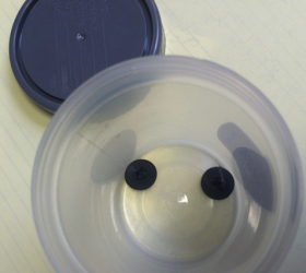
The ferret was premedicated with oxymorphone (0.1 mg/kg IM), midazolam (0.2 mg/kg IM), and glycopyrrolate (0.01 mg/kg IM). General anesthesia was induced by means of administration of ketamine (8 mg/kg IV) and midazolam (0.4 mg/kg IV). An orotracheal tube was placed and anesthesia was maintained with 2% sevoflurane in oxygen. Vital parameters monitored during anesthesia included heart rate, respiratory rate, electrocardiogram, and end-tidal CO2 using a multiparameter monitor. With the ferret positioned in sternal recumbency, esophagoscopy was performed with a pediatric bronchoscope. Two foreign bodies were visualized and retrieved with graspers (Figure 3). The objects were circular and rubbery and upon later discussion determined to be buttons from a remote-control device. Mild adjacent esophageal hyperemia was noted but esophageal examination was otherwise unremarkable. Upon completion of the procedure, anesthesia was discontinued; the ferret recovered without apparent complications and was hospitalized overnight for monitoring. Treatments included omeprazole (0.7 mg/kg PO q 24 h), sucralfate (100 mg/kg PO q 8 h), amoxicillin (20 mg/kg PO q 12 h), and syringe feedings. No abnormalities were noted overnight, and the ferret was discharged home one day post-operatively with 10 days of the medications prescribed, as well as instructions to monitor for any difficulty swallowing. Initially, the owner was advised to provide soaked kibble, with gradual reintroduction of hard kibble a week after discharge. No complications were reported, and the ferret was reportedly doing well 2 years after initial examination.
Acknowledgements: The ZCAM Department would like to thank Greylock Animal Hospital for referral of this interesting case.
* Jennifer Graham, D.V.M., Dipl. ABVP (Avian / Exotic Companion Mammal), Dipl. ACZM
Zoological Companion Animal Medicine
Department:
Foster Hospital for Small Animals