Dr. Ariana Hinckley-Boltax Publishes New Medical Illustration of Complex Suture Pattern
In her three years teaching veterinary students the intradermal suture pattern, Dr. Ariana Hinckley-Boltax (she/her) noticed another pattern—students struggling to understand it.
Assistant Professor in the Department of Comparative Pathobiology at Cummings School of Veterinary Medicine at Tufts University, Dr. Hinckley-Boltax teaches first- and second-year veterinary students in the core Clinical Skills courses.
“The intradermal suture pattern is one of the most common methods for routine abdominal skin closures, such as spays in cats and dogs. It forms a foundation of a critical surgical skill,” she explains. “Of all the suture patterns we teach, the intradermal suture pattern is one of the most complex and challenging for the students.”
The intradermal suture pattern is one way to close an incision with absorbable sutures buried in the skin, so that the pattern is not visible at the surface. Dr. Hinckley-Boltax found that students typically memorize the steps without fully grasping why each step is performed, especially the opening and closing of the suture pattern.
She found medical illustrations of the pattern lacking. “Available illustrations of the intradermal suture pattern are either poor quality, don’t show every step, or just wrong. Even some textbooks are incorrect.”
Dr. Hinckley-Boltax set out to create a new set of instructional images for the intradermal suture pattern.
“I am hoping to fill the gap in medical resources with this illustration,” she says. “This is a more conceptual approach to understanding the intradermal suture pattern and shows every step. Nothing exists to this level of depth, and it’s a needed resource for students.”
A graduate student in the Medical Illustration Master of Fine Arts program at Rochester Institute of Technology, Haley Cavarello worked as an intern with Dr. Hinkley-Boltax to generate the intradermal suture pattern images. In addition to consulting existing resources, Dr. Hinckley-Boltax would draw out or perform various aspects of the pattern for Cavarello to illustrate.
Primarily educating students in the pre-clinical phase of their studies, Dr. Hinckley-Boltax sought out the perspectives of her colleagues, who see students performing surgeries every day. She led the project in close collaboration with faculty surgeons in the Department of Clinical Sciences to ensure the images are as accurate and comprehensive as possible.
She consulted with Assistant Clinical Professor Dr. Yuki Nakayama, Assistant Teaching Professor Dr. Laurence Sawyer, Community Medicine resident Dr. Kayla Sample, Assistant Clinical Professor Dr. Jennifer Grady, Assistant Professor Dr. Mike Karlin, and Assistant Clinical Professor Dr. Kaustubh Dongaonkar. The surgeons work in the teaching hospitals at Cummings School, including Henry and Lois Foster Hospital for Small Animals and Tufts at Tech.
“It’s wonderful to have the benefit of that collaboration, it really strengthened the work,” says Dr. Hinckley-Boltax. “They are more in the weeds, they’ve done more surgeries and are exposed to more clinical experiences. They all brought something to the table in their own way. Double checking with them that I represented the surgical approach accurately was key. The panels are direct results of suggestions from Cummings School faculty.”
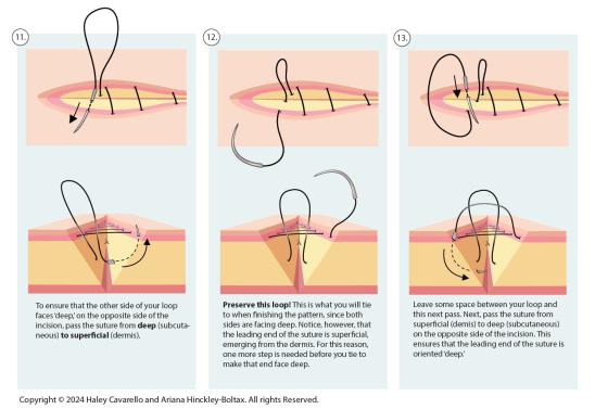
The medical illustration consists of three pages, with six panels per page detailing the intradermal suture pattern, illustrated by Cavarello with captions written by Dr. Hinckley-Boltax. Each image shows two views: the surgeon’s view and a cross-section, to closely depict every step of the procedure, since the pattern is buried in the skin and not visible from the surgeon’s vantage point.
“A lot of what is illustrated hasn’t existed before,” says Dr. Hinckley-Boltax. “It takes a conceptual approach to understanding, designed with teaching and live instruction in mind. If students understand why this complex pattern has so many steps and why it’s a good option to choose when indicated, they will be able to retain knowledge of the pattern. It goes back to educational learning theory—with adult learners, it’s really important to help them understand the ‘why.’”
The medical illustrations are already available to Cummings School’s community. Several copies have been laminated and distributed to the Joseph Kelley, D.V.M., Simulation Laboratory and other surgical spaces, including Tufts at Tech and the Luke and Lily Lerner Spay/Neuter Clinic.
Up next, Dr. Hinckley-Boltax will publish a booklet of detailed illustrations of the spay/neuter procedure in dogs and cats that she developed with another master’s student at Rochester Institute of Technology, as part of the student’s thesis.
“I have a whole list of concepts, approaches, and clinical skills that would benefit from an associated image or animation. I would like to provide the most helpful variety of resources to help students understand and perform these essential skills.”
Department:
Dept. of Comparative Pathobiology