Clinical Case Challenge: Horse with a Bladder Stone
Brady, a 20-year-old Thoroughbred gelding, was referred to Cummings School of Veterinary Medicine’s Hospital for Large Animals (HLA), to remove a bladder stone. The surgeons at HLA were tasked with determining the best course of action to remove the stone.
Can you solve the case?
History
Brady had a urolith removed at HLA a few years prior, when a small stone was obstructing his distal urethra. Due to the location of the stone, the surgery team was able to successfully remove the stone without invasive surgery to relieve the obstruction. An ultrasound and cystoscopy were performed at that time and no further stones were identified in either the urethra or bladder.
This past fall, when Brady began to urinate more frequently, Brady’s veterinarian performed an endoscopic examination of his urinary tract. This revealed the presence of a new urolith in his bladder, which led to referral back to HLA.
Determine which diagnostics and treatments are required
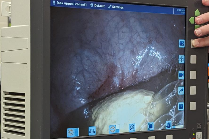
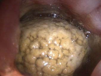
The General Surgery team started with bloodwork, which revealed normal renal function and showed no evidence of systemic inflammation. To guide treatment recommendations, Brady’s entire urinary tract— including the kidneys, ureters, bladder, and urethra—were re-evaluated. They performed an endoscopy of Brady’s urethra and bladder and confirmed the presence of a urolith in the bladder.
The Large Animal Ultrasound Service was consulted and Dr. Wade Tenney performed an ultrasound of Brady’s kidneys and ureters, observing no kidney stones or other nidus, followed by an ultrasound of the bladder. This identified the presence of an 8 cm bladder stone but no evidence of other pathology, such as a urinary tract infection or neoplasia. The ultrasound also confirmed that it was just one stone and not multiple stones within the bladder.
“We looked at the surface to see if it was smooth and shiny or spiculated with a rough appearance. We wanted to know the appearance of the stone because most are softer spiculated stones and easier to break up, but about 10 percent have a harder, smoother appearance and are more challenging to break up inside the bladder,” said Dr. Joseph Davis, Brady’s surgeon and a clinical assistant professor of large animal surgery at HLA.
Treatment: Surgery
Surgical removal of the bladder stone was recommended due to Brady’s symptoms and the risk of future complications. Dr. Davis and the surgery team considered two options for removal of the stone. The first and more traditional option was a cystotomy, which involves making an incision into the horse’s abdomen to expose the bladder and remove the stone. Although a cystotomy is a relatively routine surgery, potential disadvantages include a larger surgical incision and the need for general anesthesia, which could lead to a more difficult recovery for an older horse like Brady.
The second option was a perineal urethrostomy surgical approach using an instrument new to HLA, a pneumatic lithotripsy device. This device allows for a minimally invasive approach to be performed under standing sedation and an epidural. During the procedure, a small incision is made through the proximal urethra to access the bladder stone. A significant advantage to using this device, in addition to no general anesthesia requirement, is the stone can be broken apart in the bladder. The pieces can then be removed through a much smaller incision, resulting in less inflammation and quicker healing. Based on the size and type of stone, the team recommended proceeding with the standing procedure using the pneumatic lithotripsy device.
The surgery was a success, with no complications. This marks the first time the team performed surgery with the pneumatic lithotripsy device. Brady produced a normal stream of urine immediately after surgery. He recovered well and was discharged from the hospital two days after the procedure. He remained on antibiotics for a week and anti-inflammatory medications for five days.
The surgery site has healed, and Brady continues to fare well.
Comments from HLA’s surgical team
The most common symptom of a bladder stone is blood in the urine after exercise. Frequent urination is another indication, as is posturing with no urine production. If a urolith is suspected, veterinarians should perform a urinary tract ultrasound to confirm the presence of a stone and rule out a nidus or urolith formation. Endoscopy of the urethra and bladder is also useful in determining the presence and type of a stone, as well as evaluating the bladder for other pathologies.
In terms of prevention, urine acidifiers can help prevent stone formation. They should be used cautiously, however, as long-term use is known to cause loss of bone density in other species. Reducing calcium in a horse’s diet can also be effective, as horses naturally release calcium in their urine, and calcium can contribute to stone formation. Encouraging water intake is also important to increase urinary excretion of potential stone-forming substrates.
Determining whether to perform a traditional cystotomy or use a pneumatic lithotripsy device to remove a bladder stone is case-dependent. If it’s a larger or harder stone, anesthesia and cystotomy might be the best course of action. In other instances, the pneumatic lithotripsy device offers many advantages.
“For the most part, it’s something we can perform on a gelding standing and on mares we can do it without making an incision. In mares, we reach directly through the urethra and break up the bladder stone, so there are no wounds to heal,” explained Dr. Davis.
“With a perineal urethrostomy, we make an incision through the urethra, typically using a scope to see what we’re doing,” said Dr. Davis. “We scoop the stone into a specimen retrieval bag, pull the bag to the surface of the perineal urethrostomy site, and break up the stone through the hole right inside the bag. We then pull the chunks out of the bag where it’s all contained, so when we’re done the bladder is clean and empty, with no stone fragments left over.”
HLA’s surgical team was pleased with the outcome of the surgery and happy with Brady’s progress. “I have a specific interest in performing minimally invasive surgery. I think it’s important to offer state-of-the-art care to our clients and I’m excited about how this technology can help patients like Brady recover faster,” noted Dr. Davis.
Hospital for Large Animals
Hospital for Large Animals is a specialty hospital which boasts the largest team of board-certified specialists in one facility in New England, and a premier location to serve horses, alpacas, llamas, goats, and pigs, among others. Tufts Equine Center offers a full range of routine and advanced diagnostics and services, as well as expertise in equine pulmonary research.
Department:
Hospital for Large Animals