Advanced CT Scanner to Bring New Capabilities to Cummings School
A new computed tomography (CT) scanner is currently being installed at Cummings School of Veterinary Medicine at Tufts University. The new CT should be ready to serve patients at Henry and Lois Foster Hospital for Small Animals (FHSA) and in Hospital for Large Animals (HLA) by the middle of June. The new Canon Aquilion Exceed LB™ with Qalibra™ system will be the only CT scanner of its kind in New England with such advanced capabilities, including higher quality imaging, enhanced imaging for cardiac and respiratory CTs, standing CT scans for horses, and an enlarged bore size to scan additional areas of larger animals.
“This exciting initiative establishes Cummings School as the leading diagnostic imaging center in the Northeast, greatly expanding our clinical services and research capabilities and providing enhanced capabilities and decreased risk for our equine and small animal communities,” said Cummings School Dean Alastair E. Cribb.
CT scanners use X-ray technology to provide diagnostic imaging of the inside of the body. When Cummings School faculty determined that the current scanner was due for replacement, they explored options.
“When we have the ability to invest in new equipment that can help both large and small animals, it’s always a plus in terms of maximizing benefits to our patients, the hospitals, and the school. This new CT system can meet everyone’s needs,” said Dr. Thomas Jenei, director of Hospital for Large Animals and associate clinical professor of large animal surgery in the Department of Clinical Sciences at Cummings School.
The Canon Aquilion Exceed LB with Qalibra system will bring many advantages to HLA and FHSA. The system is made up of two separate parts working together. Dr. Jenei explains, “The Canon Aquilon Exceed LB CT scanner captures the images, and unlike human CT scans where the patient is moved in and out of the scanner, the novel Qalibra system reverses the mechanics of how we get the images.”
The new system offers a sliding gantry system that moves the CT over the patient rather than moving the patient into the CT scanner, which is critical to enable standing sedated imaging of horses. The mobile CT platform can be raised to scan a horse’s head or lowered to floor level to scan feet and fetlocks. When combined with the Canon’s large CT bore size, the Qalibra system will allow doctors to scan new areas of horses, including the lower neck, pelvis, and upper limbs.
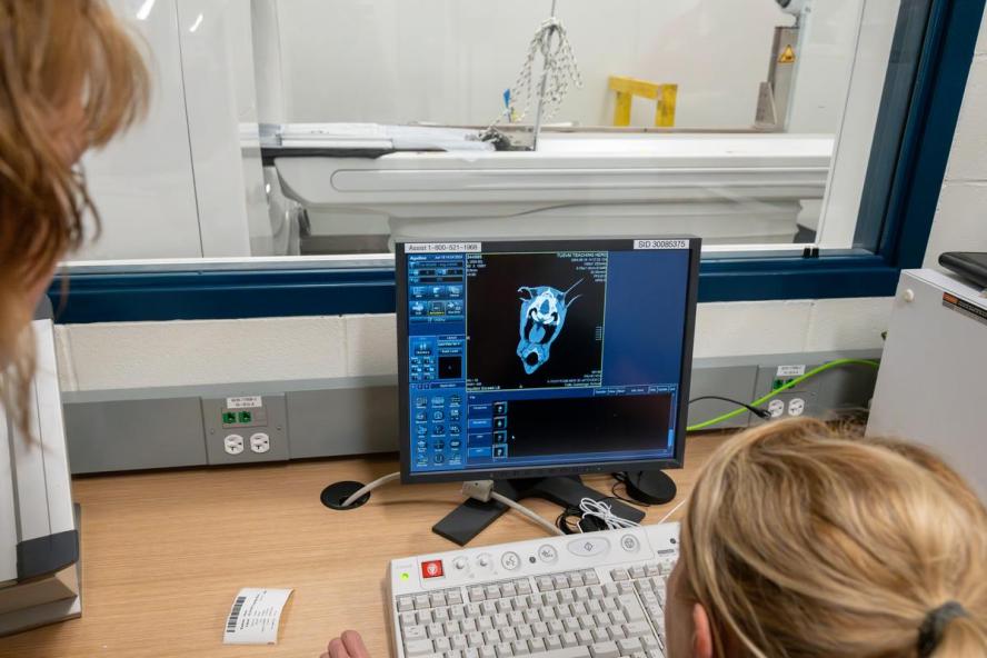
The scanner also offers high-resolution imaging, with 160 0.5 mm-thick slices to view smaller structures. This updated technology enables imaging of the smallest guinea pig patients at FHSA, horses, and other large animals from all over New England to HLA.
Standing CT scans for horses
Horses have traditionally needed general anesthesia for any CT scan, with the horse lying still on a table. For the first time, HLA will be able to perform standing CT scans for horses, eliminating the need for anesthesia in many cases.
“Anesthesia for all species brings risk, and for horses, it’s not just the risk of being under anesthesia but also waking up. There’s risk in the transition between lying down asleep and standing,” says Dr. Jenei. Horses are flight animals. Lying down feels less safe than standing. While most patients recover uneventfully from anesthesia, with heavier animals like horses, there’s a potential for injury during that process.”
Amy Sato, D.V.M., DACVR, VR00, associate clinical professor in the Department of Clinical Sciences and radiologist at both HLA and FHSA, explains that horses often present to the HLA for evaluation of fluid coming out of a nostril, which could be caused by a variety of issues such as a tooth problem or an ethmoid hematoma. These issues are best assessed via a CT scan, which would have required anesthesia using the previous CT scanner.
“Now we can scan a horse’s head while they’re sedated and standing with their head on a pillow, not under general anesthesia. It will be more convenient for owners and safer for horses because they don’t need to be knocked out completely.” In this instance, the Qalibra system will briefly move the CT scanner over the horse’s head and away again once the images are captured. Afterward, the horse can walk back to its stall and return home the same day. Anesthesia usually requires an overnight stay while recovering.
“That is very exciting to us,” says Dr. Jenei, "With the new system, we expect to be able to easily image heads and much of the legs and complete some surgical procedures with the horse standing but sedated. It’s safer and more efficient, and we can get answers faster, which is good for everyone.”
New scanning capabilities
The size of the CT scanner’s bore determines how much of an animal’s body may be scanned. HLA’s current CT scanner has a 72 cm diameter circular bore, which is too small to scan large body parts, such as the horse pelvis and lower neck. The new 90 cm bore of the Canon Exceed LB will allow doctors to scan the neck, the legs above the knees and hocks, the pelvis, and possibly even the lumbar spine of a horse.
Horses with wobbler syndrome suffer from bone pinching of the spinal cord in the neck, but the area often cannot be imaged with a traditional CT scanner because of the smaller bore. With the new system’s enlarged bore size, the team can image the entire neck to identify the affected area. “Even though, in this case, the horse would need to be anesthetized due to required positioning for larger body parts, it would allow much better assessment of horses with neck problems,” says Dr. Jenei.
Dr. Jenei explains, “For some of the expanded areas, such as the spine and pelvis, anesthesia will still be required. Previously, we could not do a CT scan of the area for conditions like wobblers, even with anesthesia. Now it will be able to be done.”
Improved image quality
The new scanner offers high resolution and more slices to view smaller structures in less time. The scans are faster, covering five times as much distance in one rotation over the previous scanner.
“This allows us to do more scans with animals sedated instead of under anesthesia for both large and small animals,” says Dr. Sato.
The new scanner also features cardiac gating technology, imaging only at specific times during a heartbeat. The motion of a heart beating can blur diagnostic images. Cardiac gating eliminates that blurring for clearer images.
“Cardiac gating is also really helpful for looking at an abnormal heart or a small tumor in the heart,” adds Dr. Sato.
Support from an anonymous donor
A foundation donated $7.5 million to acquire and install the new Canon Aquilion Exceed LB with Qalibra system. This foundation has consistently supported Cummings School in securing advanced technologies to better serve patients.
“The anonymous donor has been a tremendous supporter of the school and amazing at helping target very key needs. They are on board to support projects that are unique and special,” said Ana Alvarado, senior director of development and alumni engagement at Cummings School.
“We are very grateful for the generosity of the donor who made this possible,” says Dr. Sato. “It’s going to be a game-changer.”