Successful Removal of Skull Tumor Brings Golden Retriever Back to Health
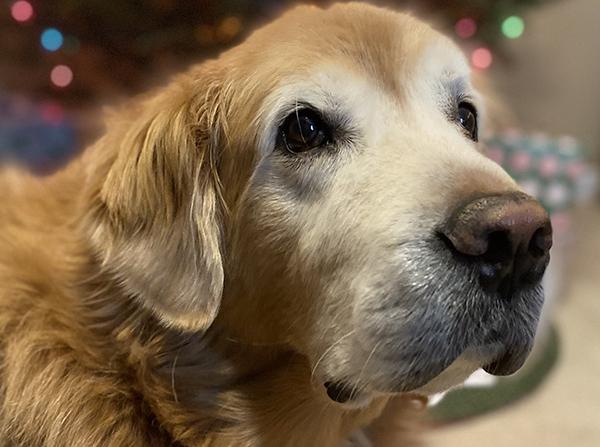
Carla and Michael Kingston noticed a bump on their Golden Retriever’s head when he was five years old. After several weeks, they grew concerned and brought Sawyer to their veterinarian, who suspected a tumor on his skull. He referred them to Foster Hospital for Small Animals at Tufts University.
A CT scan revealed a large mass covering much of Sawyer’s skull, and a biopsy confirmed the diagnosis of a multilobular tumor (MLO). Dr. Dominik Faissler, a neurosurgeon at Foster Hospital and head of the neurology residency program at Cummings School for Veterinary Medicine at Tufts, recommended surgery, radiation, or a combination of the two. The Kingstons opted for the surgery.
“Without surgery these tumors very quickly become a concern due to skull destruction and brain compression,” said Dr. Faissler. “With surgery the survival time is about one to two years because at the time of the diagnosis these tumors are already so big that complete removal with clean margins is very difficult.”
This type of surgery would be especially challenging due to tumor’s size and location.
“The tumor was compressing the brain and touching the meninges,” said Dr. Faissler. “In this situation a tumor removal with safe and clean margins is impossible.”
He explained that clean margins refer to removing the tumor and one centimeter of healthy tissue around it to eliminate all tumor cells.
“For Sawyer this would have meant that we remove a significant layer of brain tissue and the right eye,” said Dr. Faissler. “Therefore, the best option was a debulking approach with removal of all visible tumor, but not a one-centimeter rim of healthy tissue.”
Further complicating the surgery, the doctors would also need to reconstruct Sawyer’s skull. The surgery lasted three and half hours.
“We spent a lot of time carefully working around this tumor and removing all the visible mass and abnormal-looking bone,” said Dr. Faissler. “The next step was to cover the widely open brain. In Sawyer, we used the fascia of the temporal muscle and sutured a large patch over the brain. In a third step, we molded the titanium mesh to fit the skull contour and reconstructed a solid skull with a natural and physiological shape, making Sawyer look like a real dog again.”
Sawyer was treated afterwards with pain medications and antibiotics to protect the titanium implants from infection. He was released from Foster Hospital two days later in strong enough condition to walk out on his own. He continued his medications and rested for the next four weeks.
In Sawyer’s post-op appointments, Mr. Faissler was pleased by his recovery. “Sawyer appeared to be happy without neurological deficits and presented like a normal dog of his age,” he said.
The Kingstons credit the care he received at Foster Hospital for Sawyer’s healthy recovery.
“I can’t understate the value of having Tufts so close by as an option for extreme medial issues like this,” said Mr. Kingston. “The staff were incredible, not just the surgical stuff, but residents, assistants, administrators. We brought Sawyer back six months later, and the staff remembered him and all wanted to say hi.”
The success of the surgery brought the Kingstons more years with Sawyer. Mrs. Kingston was pregnant with her son Logan at the time of Sawyer’s diagnosis.
“Because of the surgery, Sawyer was able to be a part of our son’s life,” said Mrs. Kingston. She describes the two as best friends.
The tumor has not regrown in the three years since Sawyer’s surgery.
“Sawyer has reached an age of eight years and nine months, which is remarkable considering the serious medical health situation,” said Dr. Faissler.
Department:
Foster Hospital for Small Animals