It’s not every day you end up diagnosing a case that has hardly been reported in literature anywhere. In cases like this, it is the team collaborative effort that helps solve the puzzle. We’re very lucky to have really experienced and out-of-the-box thinking pathologists on both teams.
Pathology Team Pinpoints Extremely Rare Diagnosis to Save Sphynx
Severe E. coli bacterial infection documented in only four feline cases
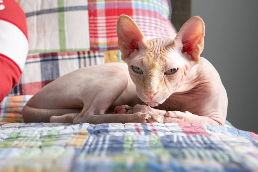
As a Sphynx, Milo’s gregarious personality comes as a surprise to most, and his recent diagnosis was just as unexpected for the clinicians and anatomic and clinical pathologists at Cummings School of Veterinary Medicine at Tufts University, who teamed together to uncover an infection that nearly took his life.
“People don’t know what to expect when they see Milo—they’re often taken aback at first but quickly realize that he’s so friendly and lovable,” says Brenda Chiaradonna of her seven-year-old Sphynx rescue. “He’s like a dog, very interactive, and loves people. He flies around the house at top speed and hangs out on top of a shelf like a gargoyle, you never know where you’re going to find him.”
A year ago last winter, Chiaradonna noticed Milo looking thin and not eating well. Their veterinarian administered fluids and an appetite stimulant. Milo ate for a few days and then stopped again. On ultrasound, the vet found his lymph nodes and spleen enlarged and recommended a follow-up at Cummings School.
By the time Milo arrived at Henry and Lois Foster Hospital for Small Animals (FHSA), he was extremely weak and had dropped a third of his body weight, down from 12 to eight pounds. The Emergency Medicine and Critical Care (ECC) team found he also had a urinary obstruction and possible intestinal mass. Feline infectious peritonitis (FIP), a disease in cats caused by coronavirus, seemed the most likely culprit, but Milo tested negative. Additional sampling was inconclusive.
Milo returned home with a feeding tube in his neck to deliver nutrients and started on a short course of steroids to treat the inflammation of his spleen and lymph nodes. Over the following weeks, he began eating on his own, put on weight, and regained his energy. His spleen and lymph nodes recovered to normal size.
The Sphynx appeared back to health until late fall when he again lost interest in food and shed pounds. On return to FHSA, his spleen and lymph nodes (even more than previously) were again inflamed, and masses were present in his intestines that looked like cancer. The lymph nodes and an intestinal mass were sampled through ultrasound-guided cytology.
Dr. Rachel Whitman, (she/her) clinical pathology resident in the Department of Comparative Pathobiology at Cummings School, conducted the initial evaluation of Milo’s cytology slides. “I saw inflammation, but it didn’t look like cancer. I suggested causes, but I was not the final stop.”
The diagnosis was still elusive. Dr. Kayla Prentice, V21 (she/her) resident in small animal internal medicine in the Department of Clinical Sciences at Cummings School, took the lead on his case. One by one, Prentice and Whitman ruled out possible conditions, from FIP to irritable bowel syndrome to less common infections, like mycobacterium. Again, he tested negative for all likely conditions.
“Kayla and I discussed typical suspects and why they didn’t add up for Milo’s case. Surgery was warranted to get a more definite answer,” says Whitman.
“Even at that point, enteroinvasive E. coli was not on our list because we usually associate that with Boxers and French Bulldogs,” says Prentice.
Prentice scheduled a surgical biopsy to obtain larger tissue samples of his lymph nodes and intestinal mass. After surgery, Milo was feeling better and returned home with appetite support (anti-nausea medications and an appetite stimulant) while they waited for the biopsy results.
Anatomic pathologist Dr. Emily Brinker (she/her), assistant clinical professor in the Department of Comparative Pathobiology, joined the case. Running histopathology on the tissue samples indicated intestinal granulomatous inflammation (or pyogranulomatous typhlitis), but the cause was still an enigma.
“The histology was consistent with a few diseases,” says Brinker. “Granulomatous colitis is a rare disease that I know of mainly in dogs. It can happen in cats; it’s very rare, but it happens. It’s our due diligence to bring up even the rare things.”
Brinker suggested more definitive testing for granulomatous colitis, a bacterial infection caused by E. coli. Prentice agreed, and they sent the sample to Cornell University’s Animal Health Diagnostic Center to run in-situ hybridization, which confirmed the presence of E. coli bacteria in the intestinal wall.
The team had a diagnosis and an unusual one—there are only four other documented cases of granulomatous colitis in cats.
“We were looking at atypical infections,” says Prentice. “Dr. Brinker’s guidance on what to do with the biopsy after the initial testing was really helpful.”
Prentice called Chiaradonna with news of the diagnosis and prescribed an antibiotic to treat the infection. That night, before she had a chance to pick up the prescription, Chiaradonna found Milo very sick on the floor and cold to the touch. She wrapped him in a heat blanket and set off for FHSA.
Milo’s blood pressure and body temperature were dangerously low. The ECC team administered heat support and intravenous fluids. Milo had the first of a number of seizures the following day.
“He was neurologically abnormal, having seizures and severely elevated ammonia levels. Having such high ammonia levels could have been a consequence of the overwhelming E. coli infection,” says Prentice. “We worried he was septic from this infection. It was hit or miss for a bit.”
The clinicians recommended a strong dose of a steroid to treat potentially overwhelming inflammation but worried about how his body would react due to the uncertainties of his possible sepsis. Chiaradonna and her husband decided to try. “I walked out thinking I would never see him again,” she recalls.
The steroid made the difference. Once stabilized, Milo began aggressive antibiotic treatment, including an intravenous antibiotic and broad-spectrum antibiotics to cover sepsis and possible infection of his central nervous system, in addition to seizure medications. He temporarily lost his vision as a result of the intensity of his seizures.
“We took it one day at a time,” says Chiaradonna. “There were days of no improvement and days where he improved a little; we held onto that. All the doctors were so kind and understanding through the rollercoaster of emotions, and very patient, explaining and helping me weigh the options.”
Milo remained at FHSA until he was healthy enough to return home the following week under the care of Chiaradonna. He continues to recover steadily and has since regained his vision. He is eating well and put back on all his lost weight.
“His little personality is coming back,” says Chiaradonna. “We’re so thankful that he has his vision. It’s almost like he’s rediscovering the world again.”
“He truly was so sick and could have died at any point,” says Prentice, who recently rechecked Milo and noted ultrasound showed his abdomen almost back to normal. “He’s doing amazing. Brenda was a trooper. We appreciated her putting her faith and trust in us. She put so much nursing care into him when he got home.”
Milo will soon taper off his seizure medications and then his antibiotics. Prentice will monitor his abdominal ultrasound, weight, and clinical signs in case of relapse.
Chiaradonna had been to FHSA once previously with her Vizsla.
“Piper got liver cancer at nine and lived to be 15 because of the surgery at Tufts,” says Chiaradonna. “I have nothing but love, praise, and thanks for everybody over there. They saved my animals’ lives. We are so fortunate to have that state-of-the-art technology and knowledge right down the road from us to help our animals.”