Imaging plays a central role
When a horse experiences a decrease in their athletic performance, it could indicate an injury or underlying disease, often best detected by imaging. Tufts Equine Center is one of the country’s select few to offer a full range of imaging services and expertise. Our team of board-certified radiologists and dedicated equine ultrasonographers rely on advanced technology to provide rapid diagnosis, treatment and recovery for equine athletes. Our clinicians work in collaboration with our on-site radiologists to interpret images in real time for a swift and accurate analysis, enabling them to solve both common and puzzling cases.
Here we review the multiple imaging modalities we use and our approach in cases.
Tufts Equine Center is fully equipped with:
- Dedicated Equine Ultrasound Suite
- Toshiba Aquilion Spiral CT Scanner
- 5 Tesla Siemens MRI
- Nuclear Scintigraphy/Bone Scanning with Siemens LFOV Gamma Camera
- Digitized Fluoroscopy with Shimadzu RS-110 Remote Machine
- Digital Radiography
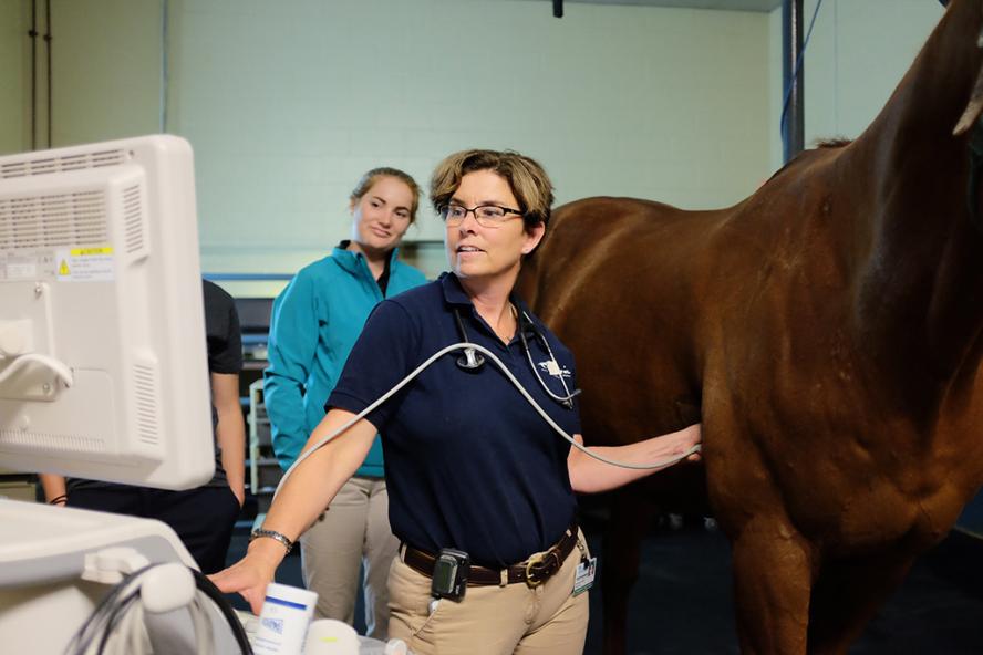
Ultrasound
Ultrasound is one of the most valuable imaging tools for evaluating, diagnosing and monitoring the healing of an injury. It is routinely used to evaluate tendons and ligaments and examine complex joints, wounds, masses and eyes, as well as the entire thorax and abdomen. This modality is highly dependent on the ultrasonographer’s experience and expertise, as they are able to detect even minute changes in bony and soft tissue structures. We use an advanced hospital-grade Toshiba Aplio 300 machine that provides high-quality, detailed images. We also have a collection of micro convex, large curvilinear, cardiac and rectal probes, which are excellent for imaging virtually every part of the horse. In addition to supporting cases within the hospital, we also offer direct consultations and outpatient ultrasound appointments. Often, we see cases where the referring veterinarian has done a thorough workup and has requested an ultrasound consultation. We take great pride in our collaborative approach to diagnosis and treatment, working hard to ensure you gain exceptional value from our partnership.
Computed Topography (CT)
CT is used most often to evaluate bony structures in more detail. It is the gold standard for imaging the equine head to detect dental, nasal and sinus disease as well as pinpointing the location of a fracture. It is also preferred for identifying tendon and ligament injuries and bone infections in foals. Because of its rapid acquisition time, it is beneficial in select cases for fracture assessment and pre-operative planning prior to undertaking repair.
Magnetic Resonance Imaging (MRI)
MRI provides the most detailed image of soft tissue structures. It takes images from numerous angles and different slices to create a 3-D image of the structure and allow for precise location of the problem. An area that is often imaged using this modality is the foot because of the machine’s ability to provide excellent images of all structures within the hoof capsule, including bones. We routinely diagnose lesions in anatomic regions such as the foot, suspensory ligament, brain and upper airway.
Nuclear Scintigraphy (Bone Scanning)
Nuclear Scintigraphy (bone scanning) is an advanced imaging modality we use to help diagnose areas of bone remodeling or active inflammation that cause lameness in a horse. This modality is routinely used when there are multiple limbs affected by lameness, or for back pain. It pinpoints areas of active inflammation so that we can focus and tailor our diagnostics accordingly.
Fluoroscopy
Fluoroscopy creates live, real-time video images of the structure inside your horse’s injured leg. Our equine surgery suites are equipped with a 9800 OEM General Electric C-Arm.
Digital Radiography (X-ray)
Digital Radiography is often the first imaging technique used to evaluate lameness by visualizing bones and joints. While portable radiographic equipment is often limited to only the legs of a horse, our high-powered unit allows our clinicians and staff to take radiographs of a horse’s chest, neck, back and head. We also rely on myelography, a radiographic examination that uses injected contrast material to detect diseases of the spinal cord.
Imaging modalities have their strengths and weaknesses depending on the situation, and we often use more than one to get an accurate diagnosis from the onset. Having the right diagnosis greatly improves your horse’s chance for a successful recovery.
Learn more about our Advanced Imaging at Tufts Equine Center