Equine Clinical Case Challenge – Clinical Signs of EOTRH
History
A 22-year old Thoroughbred gelding was evaluated for gradual weight loss and decreased appetite that had slowly gotten worse over a two-month duration. The owner reports that the gelding has otherwise been acting normal, passing normal manure and is not on any supplements or medications.
Presentation
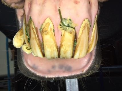
The horse was bright, alert and responsive with a body condition score (BCS) of 3/9. The last documented BCS was 4/9, 6 months ago at the time of routine vaccines. Besides being thin, the vitals and physical exam were within normal limits. The horse was observed eating and it was noted that he seemed to eat slowly and dropped a moderate amount of feed. The horse was sedated to allow for a complete oral exam and dental float if deemed necessary. The findings of the oral exam were the following: moderate points on the cheek teeth of all four quadrants and calculus, gingival recession and inflammation were noted to varying degrees on all incisors (Fig. 1).

What are your differential diagnoses?
The incisors have the characteristic appearance of equine odontoclastic tooth resorption and hypercementosis (EOTRH). EOTRH is a recently recognized disease affecting primarily the canine and incisor teeth. Characteristics of EOTRH on visual exam include gingivitis, gingival hyperplasia or recession, draining fistulas, enlargement of dental structures, tooth mobility, tooth fractures and missing teeth. Typically, a disease of older horses; clinical signs can range from asymptomatic to signs of extreme oral pain.
The differentials for weight loss and poor appetite in an older horse are numerous. Other differentials beside poor dentition which should be considered are poor nutrition, parasites, liver or kidney disease, neoplasia, etc.
What diagnostics should be performed?
Intraoral radiographs of incisors (Fig. 2): The radiographs were consistent with moderate EOTRH; obviously blunted root tips and surface irregularities are noted.
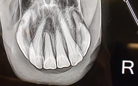
Table 1. Adjusted radiological staging system (based on Hüls et al.)
| Stage | Radiological findings | |
|---|---|---|
| Severe | 3 | Loss of tooth shape/intra‐alveolar tooth part is wider than the clinical crown/tooth fracture
Surface obviously irregular/rough |
| Moderate | 2 | Tooth shape largely preserved/intra‐alveolar tooth part is not wider than the clinical crown/obviously blunted root tip
Surface irregularly/rough |
| Mild | 1 | Tooth shape preserved/slightly blunted root tip
Surface irregularly/rough |
| Normal | 0 | No radiological findings |
CBC/Chem: within normal limits
Fecal egg count: negative
Recommendation
Since EOTRH is a painful disease and the horse was exhibiting clinical signs consistent with EOTRH, extraction of all incisors was recommended.
Outcome
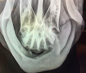
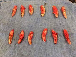
The horse was referred for extraction of all the incisors (Fig 3 and 4). Members of the American Veterinary Dental College (AVDC) Equine Dental Specialty, and American College of Veterinary Surgeons (ACVS), have received advanced training in the management of EOTRH. Following extraction, the horse was treated with 2 doses of Excede in the muscle 4 days apart and was given 3 days of phenylbutazone 1 gram twice a day post-surgery. There was some mild dehiscence but otherwise no surgical complications. Follow-up exam 3 months post-op revealed complete healing of the surgical site. The owner reports that the horse seems much more comfortable and his appetite has returned. His BCS at that time remained a 3/9 but he has not lost any more weight. Almost a year out, his BCS was back to a 4/9 and has been stable for a couple of months.
Discussion:
Equine odontoclastic tooth resorption and hypercementosis is a newly recognized disease primarily affecting equine incisors and canine teeth but has also been documented to occasionally affect cheek teeth. Named for the histological appearance of these teeth there is concurrent resorptive lesions and deposition of reparative cementum (hypercementosis). A recent study in Germany, found a 62% prevalence in horses over 10 years of age with moderate to severe EOTRH radiographical changes.
EOTRH is a painful and progressive disease. Clinical signs often go unnoticed until advanced stages of the disease. Horses may be asymptomatic or present for difficult eating, severe gingivitis, periodontal draining tracts or missing and fractured teeth. The etiology of EOTRH is still not completely understood and is likely multifactorial. Some theories that have been proposed include an immune-mediated syndrome, increased occlusal forces in ageing teeth and a potential link to bacterial infections with Treponema and Tannerella sp.
Since EOTRH is a painful disease and likely underdiagnosed disease, it is important to include examination of incisors and canine teeth in every routine dental exam and float. If changes consistent with EOTRH are noted, intraoral radiographs should be recommended. There is currently no proven medical management of EOTRH and horses with clinical signs should be considered candidates for extractions to improve overall health and quality of life.
References:
- Smedley R.C., Earley E.T., Galloway S.S. et al. Equine Odontoclastic Tooth Resorption and Hypercementosis: Histopathologic Features. Vet Pathol .2015;52: 903-909.
- Lorello O., Foster D.L., Levine D.G., et al. Clinical treatment and prognosis of equine odontoclastic tooth resorption and hypercementosis. Equine Vet J. 2016; 48: 188-194.
- Hole S.L and Staszyk C. Equine odontoclastic tooth resorption and hypercementosis. Equine Vet Ed. 2018; 30: 386-391.
- Rehrl, S., Schroder, W., Muller, C. et al. Radiological prevalence of equine odontoclastic tooth resorption and hypercementosis. Equine Vet J. 2017; 50: 481-487.
- Sykora, S., Pieber, K, Simofer, H. et al. Isolation of Treponema and Tannerella spp. From equine odontoclastic tooth resorption and hypercementosis related periodontal disease. Equine Vet J. 2014; 46: 358-363.