Clinical Case Challenge: More Than Hip Dysplasia?
Can You Solve the Case of the Month?
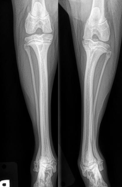
A recently adopted eight-month-old male Shetland sheepdog was walking oddly. His family also noticed his left hind limb appeared crooked. His owners brought him to their primary veterinarian, radiographs were taken, and he was initially diagnosed with hip dysplasia and a limb deformity. (See Image 1)
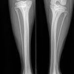
Over the next two months, the left hind limb deformity worsened, and a follow-up appointment was scheduled with the primary care veterinarian. During the follow-up visit, the patient was also diagnosed with a left hind leg grade 3 medial patella luxation and increased valgus deformity of the distal tibia. Radiographs of the coxofemoral joints, the left stifle, and the left distal tibia revealed mild evidence of hip laxity, medially luxated left patella and suspected damage to the distal lateral tibia growth plate. The medical team determined, the best course of action would be to compare the right tibia to the left tibia.
Q. What are your concerns and how would you proceed?
A. Concerns: given his age and having an active growth plate surgical correction could be complicated. Next steps: perform CT scan for surgical planning.
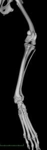
The CT scan confirmed the physical examination and radiographic findings. A 3-D reconstruction was recommended and performed. The medical team completed surgical planning with measurements for angular limb deformity. (See Image 2)
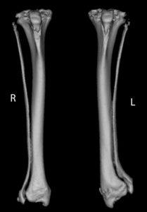
The patient was taken to surgery where a medial closing wedge of the distal tibia was performed to correct the limb deformity. The trochlear groove was checked and was noted to be of adequate depth. However, the patella was wider than the groove, so each edge of the patella was trimmed/narrowed enough to ensure it fit within the groove. (See Image 3)
The patient did well post-operatively and stayed in New England until his two-week recheck. After the recheck, he and his family returned to Florida to enjoy the warm weather for the winter. He is doing well and enjoying the sun and warmth of the south. Radiographs were taken 12 weeks post-surgery and showed adequate healing.
Angular limb deformities can be the result of many different factors and are often best treated with a surgical realignment of the axis of the bone.