Our ideal goal is to change the paradigm of using the mouse as the primary model for this disease. Some of the research conducted over the last two to three decades, using the mouse model, did not necessarily provide data that’s really reflective of natural infections in humans.
Reinventing the classic animal model
Cummings School research team uses guinea pig model to advance tick-borne disease research
Scientists who study human disease processes often go to great lengths to find an ideal animal model that recapitulates natural disease. Working with vector-borne diseases adds another challenge because the vector, in this case a tick, creates a unique environment during transmission that may affect disease development.
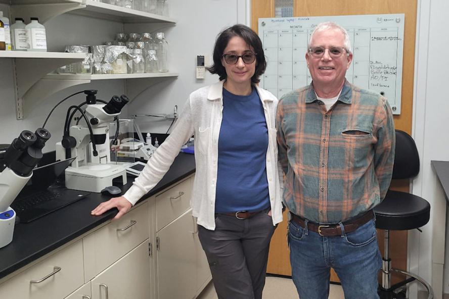
One pair of scholars united nearly 20 years ago to study tick-borne diseases. Dr. Andrea Varela-Stokes, V01, is professor and chair of the Cummings School of Veterinary Medicine Department of Comparative Pathobiology. Her co-investigator and spouse of over 19 years, John Stokes, is also Cummings School’s director of the new Flow Cytometry Shared Resources.
They met as graduate students at the University of Georgia, where Varela-Stokes earned a Ph.D. and Stokes an M.S. Though they initially worked together on a Lyme-like illness in the South (Southern Tick-Associated Rash Illness), Stokes ultimately joined a malaria lab for his thesis work. Nonetheless, they soon discovered how effective they were as a team and that having each other constantly challenge ideas could lead to something better than they could do separately. That and their respective strengths, including working with animal models, infectious diseases, and immunology, “created a synergy and has been the foundation of our scientific partnership,” says Varela-Stokes.
Varela-Stokes and Stokes worked on various tick-borne disease projects “off the record” for nearly a decade. Their formal collaboration started in 2014 at Mississippi State University, where Varela-Stokes was on faculty before joining Cummings School and had been primarily studying tick-borne infection dynamics in reservoir hosts, both vertebrate and invertebrate.
Stokes joined as co-investigator after getting the University’s flow cytometry shared resource started, and they redirected their research to take advantage of his immunology expertise – pivoting to studying the disease process in spotted fever rickettsiosis. Since then, they have collaborated on numerous funded projects, published 15 papers (so far), and are raising their son, who just started high school. The team joined Cummings School in March 2022.
Their spotted fever rickettsioses research
Spotted fever rickettsioses comprise several diseases ranging from mild to deadly, the deadliest being Rocky Mountain spotted fever (only studied in Biosafety Level 3 facilities). The agents, spotted fever group Rickettsia species, are transmitted by various tick species. To ease the inclusion of student researchers at various levels of their training – from undergraduates to graduate students – the team uses a less pathogenic agent, Rickettsia parkeri, which was first associated with human disease in the early 2000s. “We look at it from the disease standpoint, using a guinea pig model and natural transmission,” said Varela-Stokes—so, using tick feeding on the guinea pigs to transmit the pathogen, as would occur in humans.
Both researchers strongly advocate using a guinea pig model, which acquires the disease naturally and has an immune system more like humans than mice do. “The mouse became the primary model in the late 1980s after the development of genetically modified mice,” Stokes shares. “With an ability to facilitate genetic manipulations and because it’s the less expensive option, most researchers adopted the mouse model.”
In the early 1900s, when Rocky Mountain spotted fever was first identified, guinea pigs were used to help identify the agent and for much of the subsequent research, Varela-Stokes says. They were ideal models then—for demonstrating the disease, being easy to handle, manageable when tick-feeding, and large enough to collect many samples at multiple time points during disease progression. The team purported that it was time to return to that model for the same reasons, and considering no mouse model had those advantages.
Using Stoke’s technical expertise in molecular and immunoassay design, they started to develop the tools and assays necessary to study the disease in the guinea pig.
Research published in Current Protocols
Their most recent paper, “Evaluating the Clinical and Immune Responses to Spotted Fever Rickettsioses in the Guinea-Pig-Tick-Rickettsia System,” was published in the Immunology section of Current Protocols e584, Volume 2. Stokes served as lead author, in collaboration with Varela-Stokes as senior author, with co-authors including their previous team at Mississippi State University and colleagues at the Centers for Disease Control and Prevention (CDC).
“This paper combines many of the assays that we’ve developed along with some that others have done at CDC,” Varela-Stokes explains. “[The CDC’s] Michael Levin is on that paper. He’s worked a lot with guinea pigs and supports that model. Sometimes we run into some barriers with folks who would like to stay with their traditional mouse model.”
Overall, they are building the picture of the disease within the guinea pig because it best replicates the clinical signs seen in humans exposed to Rickettsia through tick transmission. By developing an enzyme-linked immunosorbent assay (ELISA) for detecting guinea pig antibody levels to rickettsiae, they developed an alternative to the more subjective microscopic assays (i.e., immunofluorescent antibody tests or IFA) used by most diagnostic labs. Ultimately, they hope to adapt this to distinguish exposure to different spotted fever group Rickettsia species in humans, which cannot be done using the IFA.
Using flow cytometry, they can quickly differentiate many immune cell types circulating in guinea pig blood before and after infection; they recently expanded that to identify the immune cells that infiltrate the skin when ticks transmit the rickettsiae. The next step is to identify the cells infected with rickettsiae using flow cytometry. There is much work left to do, though, as they acknowledge that the limited number of reagents forces them to become more creative and determined in their efforts.
Changing the paradigm to use guinea pig model for this research
“Our ideal goal is to change the paradigm of using the mouse as the primary model for this disease,” Varela-Stokes shares. “Some of the research conducted over the last two to three decades, using the mouse model, did not necessarily provide data that’s really reflective of natural infections in humans.”
Stokes and Varela-Stokes are revisiting some of those studies with a guinea pig model and extending the studies further. “Part of that may confirm some of the mouse data … but the hope is that it will provide a greater level of confidence in the data that we require for our understanding of spotted fever rickettsioses in humans,” Varela-Stokes explains.
They are also excited about the potential for detecting infection early using biomarkers in the guinea pig model. Ultimately, they believe that detecting infection early and teasing apart the mechanisms underlying the development of spotted fever rickettsiosis after natural tick transmission is best met using the guinea pig model.
Department:
Dept. of Comparative Pathobiology