-
About
- Leadership & Faculty
- News & Events
-
Admissions
-
Academics
- Graduate
- Advanced Clinical Training
- Continuing Education
- Academic Departments
- Academic Offices
- Simulation Experiences
-
Student Life
- Offices
-
Research
-
- Transformative Research
- Centers & Shared Resources
-
-
Hospitals & Clinics
- Emergency Care
- Hospital Services
-
Community Outreach
- Volunteer
The Hunt for a Porcupine Quill Saves Dog from Losing Eye
Foster Hospital’s Diagnostic Imaging and Ophthalmology Teams work together to locate and remove migrating quill

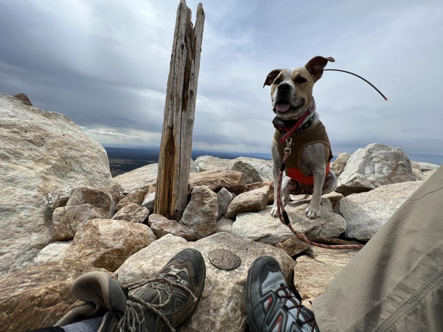
Hiking on a mountain trail near their home this past spring, Nicholas Pignatello, Danielle Sanville, and Lucy, their 10-year-old Pit Bull Terrier and American Staffordshire Terrier mix, happened to cross paths with a porcupine on their descent. Lucy approached the porcupine and was soon covered in hundreds of quills.
Still an hour’s hike from the trailhead, they could not carry Lucy due to the quills protruding from her face, chest, and shoulders, so she walked down on her own, quills and all.
“Every time we stopped, she pawed at her face, so we tried to keep her moving and then took her straight to the ER,” recalls Pignatello.
Pignatello and Sanville rescued Lucy when she was about a year old. “I think she rescued us. She comes everywhere with us. We travel all over and always bring her with us,” says Sanville, who describes Lucy as a sweet, friendly dog that loves balls and snuggling.
The doctors at South Deerfield Veterinary Clinic removed all the quills they could find that day—and over three subsequent visits, as the quills migrated to other parts of Lucy’s body, including on the back of her neck, around her eye, and even on her rib cage opposite to where she was hit.
Two weeks after the initial encounter, Lucy’s eye swelled, with redness and tearing, her bottom eyelid especially irritated. The veterinarians at South Deerfield suspected another quill but could not locate one or an alternate source of the injury, so they referred Lucy to Henry and Lois Foster Hospital for Small Animals (FHSA) at Cummings School of Veterinary Medicine at Tufts University.
“The tricky thing about porcupine quills is that they can break off and migrate to areas they shouldn’t and cause trouble,” says Dr. Stephanie Pumphrey (she/her), assistant professor of veterinary ophthalmology in the Department of Clinical Sciences at Cummings School and medical director of specialty services at FHSA. “We were fairly suspicious that a quill was in there.”
A common injury for dogs after contacting a porcupine, the quills enter the dog’s tissue and break off. Locating migrating quills can be difficult. The Ophthalmology Team pulled in the Diagnostic Imaging Team at FHSA to help search for the suspected quill.
“Porcupine quills present a diagnostic challenge—they are essentially invisible on X-ray and CT scans. We really can’t see quills confidentially on any modality other than ultrasound,” says Dr. Nathan Biedak, diagnostic imaging resident in the Department of Clinical Sciences at Cummings School. He explains that ultrasounds cover a much smaller body area than a CT scan or MRI.
Three porcupine quills appeared on Lucy’s ultrasound, two superficial and one fragmented and fractured, measuring approximately one millimeter in thickness and two centimeters long, just below the back of the eye.
“One of the biggest problems was that it was very close to contacting the eye itself. It was behind the eye, with less than half a millimeter from going into the globe. We couldn’t confidently say if it was penetrating the eye, but it was very close,” says Dr. Biedak. “I’ve seen other quills in the past, but not in this area. This was my first quill hunt around the eye.”
Dr. Pumphrey recommended surgery as the best option. The Ophthalmology Team hoped to spare Lucy’s eye, but if the quill were actually in the eye or not in an accessible location, they would have to take out her eye.
Surgery was scheduled for that afternoon. The Diagnostic Imaging Team advised the Ophthalmology Team on the exact location of the quill.
“We work with the Diagnostic Imaging Team fairly frequently—this was an unusual case because of the quill aspect,” says Dr. Pumphrey. “We talk with them to determine what imaging is best for the patient and how we will get everything done. They provide guidance about how to treat during surgery and how deep to go. They are great partners and make our jobs a lot easier.”
Based on the Diagnostic Imaging Team’s guidance, Dr. Pumphrey and her team dissected carefully around Lucy’s eye and found the quill next to the eye. Fortunately, the quill had not perforated the eye itself. They removed the quill without having to take her eye and without causing any damage to her eye. They also removed the additional quills near her eye.
“The ultrasound results didn’t seem too promising in terms of saving her eye. One quill was directly behind her eye socket, pointed at the eye. We were told there was a pretty good chance she might have to lose her eye,” recounts Pignatello. “A couple hours later, we got the call that they could save the eye, which was a huge sigh of relief. Everyone was very excited. It was a big moment, and we are thankful for it.”
After the surgery, Dr. Pumphrey showed the tiny quill to Pignatello, Sanville, and the Diagnostic Imaging Team, who were all thrilled at the results.
“It was so impressive that the surgeon came out with the quill to talk with us. She said she and her team were ecstatic to save the eyeball. We were, too,” says Sanville.
Lucy was in and out of FHSA the same day she arrived and went home with the quill. A few weeks later, she returned to have her stitches removed and to check that she didn’t have any more lingering quills.
“Lucy is a nice example of a great dog with great owners and services working together in the hospital to get a good outcome for the dog,” says Dr. Pumphrey.
Sanville and Pignatello report that Lucy fully recovered and is back to hiking.
“It was an amazing experience at Foster,” says Sanville. “Everyone there was unbelievable—friendly and attentive. The facilities and entire staff were top-notch. It was one of the best experiences we’ve ever had going to an ER vet. Driving in there, you know you’re headed for something special.”
Department:
Foster Hospital for Small Animals