Equine Clinical Case Challenge: Arrhythmia in a Show Jumper
History:
A 17 year- old warmblood show jumper was evaluated on the farm for mild exercise intolerance and prolonged recovery after exercise, which was present despite a decrease in active competition level from upper level to lower level jumper. An irregular rhythm was detected, and the warmblood was referred to Tufts Equine Center at Cummings Veterinary Medical Center for cardiac evaluation.
What would be your next step during the examination?
Presentation:
Auscultation revealed an irregular rhythm, heart rate of 36 bpm, and a left sided Grade 2/6 soft blowing early systolic murmur. Mucous membranes, CRT, arterial pulse quality, respiratory rate and effort were within normal limits. Further questioning indicated that the horse’s last known normal auscultation was at least 6 months prior.
What are your differential diagnoses?
An irregular rhythm is the hallmark of atrial fibrillation. Atrial fibrillation is the most common performance affecting arrhythmia in the horse. Typically, it has the most effect on horses performing at a high level of cardiovascular requirement; however, the degree of exercise tolerance varies amongst individual animals.
Differentials for a left sided Grade 2/6 holosystolic murmur in an otherwise normal adult horse include physiologic flow murmur or mitral valvular regurgitation. Physiologic murmurs are typically low grade (<3/6) murmurs that change in grade or disappear with changes in heart rate. Echocardiographic evaluation should help differentiate and can quantify severity of any disease present.
Lower airway or other respiratory disease should be considered and ruled out in any horse presenting for exercise intolerance /prolonged recovery, with or without concomitant cardio-vascular disease.
What diagnostics should be performed?
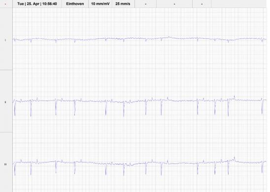
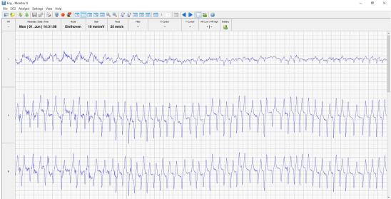
ECG (Fig1): The resting ECG confirmed atrial fibrillation.
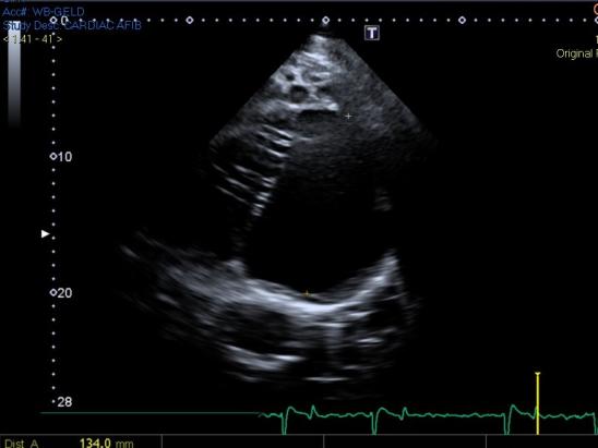
Echocardiogram (Figure 2 and 3): The echocardiogram revealed mild mitral regurgitation (Fig.2) with no structural valvular pathology and a left atrial size on the upper end of normal (Fig.3) It is particularly important to evaluate any potential valvular regurgitations and determine left atrial size in horses with atrial fibrillation as mitral regurgitation and left atrial enlargement are risk factors for recurrence. Horses with significant cardiac disease are not considered candidates for conversion.
Fig. 2 below
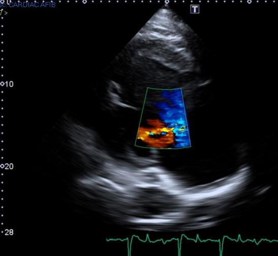
Fig. 3 below
Exercising ECG (Fig. 4): Radiotelemetric heart monitors can be used during exercise to determine if the heart rate during is appropriate for each level of exercise and if any ventricular ectopy is occurring. The gelding demonstrated a heart rate of up to 240 bpm at a fast trot and slow canter with two short runs of ventricular tachycardia prior to discontinuation of the test.
Lung function testing: to be considered pending cardiac result, given lack of evidence for infectious respiratory disease or lower airway disease.
CBC/CS: Within normal limits
Fractional Excretion of potassium: Within normal limits
CTnI: within normal limits
Recommendations: Conversion to normal sinus rhythm is recommended when the average maximal heart rate during exercise at an intensity that is at or slightly exceeding the horse’s normal activities is greater than 220/min, signs of exercise intolerance are present and/or all horses undergoing rigorous work. The presence of ventricular ectopy raises concerns for the development of unstable ventricular arrythmias which could lead to collapse or sudden cardiac death. Although duration of atrial fibrillation may have been up to six months, which might result in less likelihood of successful conversion and maintenance of normal sinus rhythm, attempt at conversion was the best chance for him to be usable as a show horse.
Outcome: The gelding was successfully converted to normal sinus rhythm following the four doses of quinidine sulfate q2 hours per nasogastric tube. Transient diarrhea and paraphimosis were noted and resolved uneventfully within 12 hours after conversion. Post conversion echocardiogram was performed and 24 -hour holter monitor recommended to assess for atrial or ventricular ectopy. He was discharged with recommendations for four-six weeks of rest given the unknown duration of his atrial fibrillation and resulting amount of atrial remodeling, and his ventricular ectopy. Periodic auscultation was recommended to monitor for recurrence. The horse returned successfully to a career as a lower level jumper without reported recurrence for one year to being retired as a competition horse. Current rhythm is unknown.
Discussion: Atrial fibrillation can occur secondary to cardiac disease or occur in isolation (lone atrial fibrillation). It can be paroxysmal or sustained. In paroxysmal atrial fibrillation, spontaneous conversion to normal sinus rhythm occurs typically in under 48 hours. Horses with lone atrial fibrillation typically have normal resting heart rates and no outward clinical signs at rest or low-level exercise. During exercise, horses in atrial fibrillation experience heart rates of 20-40 beats per minute higher than a horse in normal sinus rhythm in order to cope with the decrease in cardiac output the condition entails. At lower levels, up to even submaximal exercise levels (<180 bpm) this may not result in a change in performance. However, those working near maximal cardiac output (such as racehorses, 220 bpm) or those individuals having inappropriately high ventricular response rates while in atrial fibrillation will have performance limitation. Collapse during exercise has been reported in horses with atrial fibrillation. More recently, in groups of horses with atrial fibrillation and exercise intolerance, ventricular arrhythmias have been documented even at relatively low levels of exercise. Exercise tests using radiotelemetric heart monitors can be now performed under a variety of conditions in the field. It is recommended to assess the ECG under conditions which most simulate their use in order to best approximate workload and stimulate normal vagal responses.
Conversion options include treatment with oral quinidine sulfate or transvenous eletro cardioversion. Success rates of 65-90% are reported with both treatment types, with the highest success rates typically seen in lone atrial fibrillation of short duration. Recurrence rate is to 40% overall; recurrence rates of lone AF and less than one- month duration is about 15%. Horses with chronic valvular disease, atrial enlargement, excessive atrial premature contractions and/or previous unsuccessful conversion attempt have more likelihood of recurrence. Longstanding duration of atrial fibrillation (>4 months) results in time dependent electrical and structural remodeling and is considered to decrease chances for successful conversion and increase likelihood of recurrence. Conversion is not recommended if significant cardiac disease is present. Ridden or driven horses in whom conversion is not an option should have yearly exercise testing performed and should be ridden by informed consenting adults only.
Dr. Kate Chope is a lifelong horse owner and competitor, having successfully shown through Grand Prix in show jumping. She is also one of the few academically-trained veterinary ultrasonographers specializing exclusively in equine ultrasound. Dr. Chope is board-certified by the American College of Veterinary Sports Medicine and Rehabilitation and brings strong expertise in clinical ultrasound in her work as a clinical faculty member at Cummings School of Veterinary Medicine at Tufts University. Dr. Chope earned her VMD from the University of Pennsylvania, where she also undertook a fellowship in equine cardiology, served as an academic lecturer and has contributed to several clinical articles in peer-reviewed veterinary journals.
References:
- Reef VB, Bonagura J, Buhl R,et al. Recommendations for Management of Equine Athletes with Cardiovascular Abnormalities: Joint ACVIM/ECEIM consensus statement. J Vet Intern Med 2014; 28:749-761
- Verheyen T, Decloedt A, van Der Vekens N et al. Ventricular response during lungeing exercise in horses with lone atrial fibrillation. Equine Vet J 2013; 45: 309-313.
- Decloedt A, Schwarzwald C, De clercq D et al. Risk factors for recurrence of atrial fibrillation in horses after conversion to sinus rhythm. J Vet Intern Med 2015; 29: 946-953
- Allen K, Young L and Franklin H. Evaluation of Heart Rate and rhythm during exercise. Equine Vet Educ.2016:28 (2) 99-112