Clydesdale-Thoroughbred Cross Undergoes Successful Corneal Transplant at HLA
A Clydesdale Thoroughbred cross named Jax (short for Sir Jackson) and his owner Stephanie Juriansz have quite a bit of fun together. They compete in dressage, show jumping, and cross-country events and enjoy fox hunts and trail rides. When Jax was on stall rest for a month with an injury, Juriansz taught him a few tricks—he can bow, dance on command, retrieve a tossed glove or crop, and lift his lip to smile. And he's addicted to Twizzlers.
"He's just this big, big horse with a lot of personality. He loves trails and always lets out a little squeal when he goes running," says Juriansz.
Seventeen hands tall, Jax lives in a pasture on a small farm with three other mares and a gelding in Lincoln, Massachusetts. Late last summer, his right eye began to swell. Their veterinarian came out to the farm and thought it might be an allergic reaction. They applied topical steroids to the eye, but it worsened over the next several days. The vet recommended he see an ophthalmologist.
Juriansz brought Jax to Hospital for Large Animals (HLA) at Cummings School of Veterinary Medicine at Tufts University. HLA's Ophthalmology service diagnosed him with a corneal ulcer, an open wound on the cornea. He had both fungal and bacterial infections in his cornea, diagnosed from a swab taken from the eye.
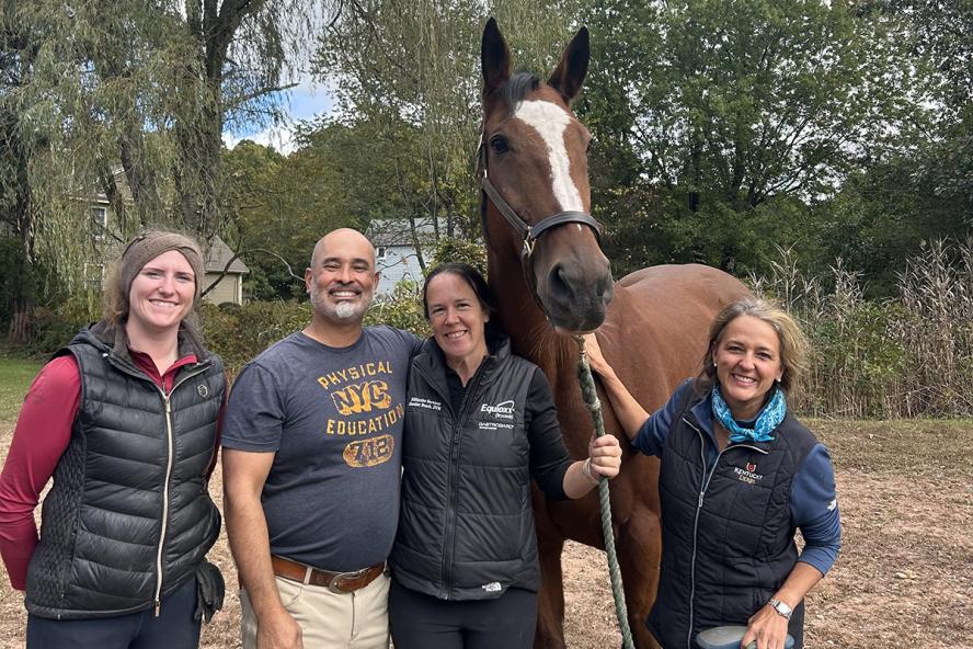
"He had a mixed infection of the cornea. In horses, it's not uncommon for them to get both fungus and bacteria because they live outdoors," says Dr. Vanessa Yang, on the Ophthalmology team at HLA and Henry and Lois Foster Hospital for Small Animals, and assistant professor in the Department of Clinical Sciences at Cummings School.
Jax likely had a small trauma to his eye, such as being poked by a piece of hay, allowing the bacteria and fungus to take hold. The Ophthalmology team placed a subpalpebral lavage line (SPL, similar to a catheter) in his eyelid to administer medications without having to touch sensitive areas of Jax's face, since corneal ulcers can be painful. He was given more than a dozen eyedrops a day through the lavage system, including antifungals, antibiotics, and others.
Within a few days, the ulcer sealed itself over, but a stromal abscess had formed within his cornea. The abscess was large, measuring more than six millimeters. Stromal abscesses in horses are a vision-threatening disease and may lead to loss of the eye in some cases. So, in addition to the eyedrops, the team performed intrastromal injections of an antifungal medication into his cornea. After weeks of aggressive medical management, the abscess was not healing.
"When they develop these deep stromal abscesses, they're more difficult to treat than superficial ulcers," says Yang.
With the severity of the case and without seeing any progress, surgery was the next viable option for treatment.
"You could tell he was very irritated and uncomfortable, and I really wanted to give him a chance," says Juriansz. "It was a hard decision. The Tufts [Cummings School] team is so professional. Dr. Yang was very patient and explained everything thoughtfully. At the end of the day, I'm just so grateful I made this decision. It may have taken a different course if I had waited too long to respond."
A penetrating keratoplasty, a type of corneal graft, involves removing the area of infection from the cornea and transplanting healthy corneal donor tissues. Yang has performed corneal transplants in horses, dogs, and cats.
"Because the abscess is a very focused, discrete area of infection and the surrounding cornea is healthy, we can essentially cut out that area of infection," explains Yang. "We placed a corneal transplant within the defect, sutured that in, and we overlaid that with a second graft called a conjunctival graft, which is made of Jax's own tissue."
Covering the donor tissue with Jax's tissue brings blood vessels and accelerates the healing process.
"This is a good example of us trying a couple of different things before we had to reach for surgery ultimately," says Yang. "It's a lot of teamwork to put these horses under general anesthesia, so when we do procedures like this, we loop in the Large Animal Surgery team, who help us with induction into anesthesia and recovery. Large Animal Internal Medicine also helps us with these cases. He had some mild complications from his oral anti-inflammatories, which helped us manage that. It was definitely a combined effort between the Ophthalmology service and everybody else at Hospital for Large Animals."
After recovering for a few days at HLA, Jax moved to a rehabilitation facility in Connecticut. He continued his medications through the lavage system, and the rehabilitation team stayed in close contact with Yang throughout his recovery.
"Jax was a really good boy; he was good for his treatments," says Yang. "Stephanie visited him all the time and always refilled his Twizzlers when he ran low."
Jax returned to his herd on the farm in October.
"He's happy, running around, and back to himself," says Juriansz. "I'm grateful for the Tufts team, I'm grateful for the rehab team, I'm grateful for my vet, who came out right away. She tried her best and knew it was time to kick it up to Tufts. I'm feeling so blessed that Jax is stable, and he has his vision."