Model development is really interesting because it takes using prior research, using real anatomy, and trying to figure out how to model that using different materials for better learning opportunities. I've very much enjoyed my experience. It's been nice to have this summer where I can be all in on this and do it to the best of my abilities.
Student Summer Researcher: Jakob Johnson, V28
Johnson designed a new training model for the Sim Lab to test intraocular pressure

The Joseph Kelley, D.V.M. Simulation Laboratory (Simulation “Sim” Lab) will have a new model created by Jakob Johnson, V28 (he/him), a student in the Student Summer Research Program at Cummings School of Veterinary Medicine at Tufts University. He developed an eyeball model for students and residents to practice measuring intraocular pressure.
Johnson’s research, “Project Development and Evaluation of Silicone-Based Canine Eye Models for Intraocular Pressure Measurement Using TonoVet Rebound Tonometry,” was selected as a first-place award winner in the category of Clinical and Translational Research at the 37th Annual Veterinary Research Day on September 11.
Dr. Mike Karlin (he/him), director of the Simulation Lab and associate professor in the Department of Small Animal Clinical Sciences at Cummings School, was Johnson’s advisor for the project. “Jake knocked it out of the park. I'm so impressed. He put his head down, got the job done, and came out with a model that was 10 times better than I expected.”
Johnson’s first research experience was as an undergraduate neuroscience major participating in a study investigating the effects of feed change on methane gas emission from cows and later a retrospective study on Parkinson’s disease. Johnson has recently become interested in ophthalmology and orthopedics. He connected with Karlin, who helped start the Sim Lab and has been integral to developing new models for students to practice skills before a live setting.
Students designed many of the models in the Sim Lab, including the very first model of a simulated spay abdomen. The lab does not yet have an eyeball model to practice gauging intraocular pressure to diagnose conditions such as glaucoma and uveitis. The goal of Johnson’s research project was to determine if silicone could be used as a material to create an eyeball model to measure intraocular pressure.
“A realistic model would allow students in training to be able to hone their skills for measurement on eyeballs, so that when they actually have a patient in front of them, they've had the experience to do the procedure,” says Johnson. “We weren't sure how we were going to approach it to start, so that was exciting because it was more trial and error.”
Johnson tested 22 different eyeball prototypes before landing on three that could potentially work. The initial solid silicone model was not effective, so he pivoted to creating a silicone cast of an eyeball that could be filled with fluid. Johnson relocated to the Sheep Barn to utilize the rotocaster, a rotating casting machine that spins the silicone until it covers the inside of the mold and forms the shell of the model eye. He then attached a tube to the model, sucked out the air, and injected fluid to set the model to the necessary pressure.
“At the beginning of the summer, a lot of it was starting from scratch for me—figuring out model making and how to use 3D design programs,” says Johnson. “Dr. Karlin was super helpful in assisting me and a good sounding board to bounce ideas off of. He was always available to help out, but also gave me freedom to try stuff out, maybe fail, and then figure out what worked from there, which was valuable for me, instead of telling me what he thought should be done right away.”
After creating a hollowed-out silicone shell, Johnson found water worked better than pressure gels to gauge pressure. He could not identify one silicone material that spanned low, normal, and high pressure ranges, so he created two models.
“We put one model in one eye socket that would cover the high-end range, and then one in the other eye socket that can adjust between a low pressure and a normal pressure. Based on how much water you inject in, you can adjust the pressure,” he says.
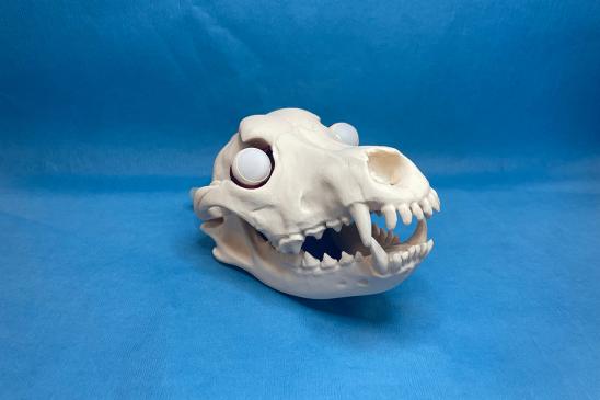
Johnson faced the additional challenge of creating a physiologically realistic skull to house the eyeballs. He spent the last month of the project on developing the skull model, and when he landed on a viable design, he printed the skull in a bone color and the muscles in red. A tube through the back of the skull inserts into the eyeball models to inject the amount of water needed to test different pressures.
One of the first skull prototypes did not look realistic enough for Johnson, who wanted it to appear more natural. “We were flip-flopping between how realistic versus how practical it needs to be for learning purposes. I did a lot of research and used a lot of materials to figure out positioning and anatomy of the muscles that hold the eye in the orbit. I designed all of the extraocular muscles and a little cup that holds the eyeball.”
“Jake did an amazing job,” says Karlin. “The model has the ability to help improve students’ confidence, so that they go into clinics more comfortable performing the procedure on a live patient after practicing on the model.”
Cummings School’s ophthalmology faculty found Johnson’s model to be an accurate representation of the eye and reported it performed well in measuring intraocular pressure. The model will be incorporated into the ophthalmology lab in the Clinical Skills course.
“Originally the project was to create this model for research purposes. It's pretty rewarding that they're going to use this in the future,” says Johnson.
The model is so realistic and practical that Johnson and Karlin are considering uses beyond testing intraocular pressure, such as practicing nerve blocks or adding a function to view the back of the eyeball to evaluate different retinal pathologies.
“We didn't expect it in the beginning, but it's opened our eyes to where this could go with certain other ophthalmologic procedures, injections, or taking fluids from the eyeball, which are more specialized and require a lot of practice,” says Johnson.
Johnson says of his summer research experience, “Model development is really interesting because it takes using prior research, using real anatomy, and trying to figure out how to model that using different materials for better learning opportunities. I've very much enjoyed my experience. It's been nice to have this summer where I can be all in on this and do it to the best of my abilities.”