Heart Disease Diagnosis
Heart Disease Resources: Basics | Symptoms | Diagnosis | Dogs | Cats | Treatments | Medications | Nutrition | Forms
Diagnosing a dog or cat with heart disease can be quite the journey. Some pets are diagnosed with a heart condition based on findings from the physical exam. Other pets may require extensive testing to determine the cause of their issues. Testing will likely be required to understand the intricacies of your pet’s heart condition.
A visit to the veterinarian for possible heart disease will begin with a detailed discussion. Your veterinarian will want to know about the history of the problem and any treatment. It will help if you can tell them about the frequency and duration of any symptoms (See our Symptoms page). If your pet is taking any medications be sure to bring the medications. If you have kept track of breathing rate and effort (Monitoring form), or if you have completed one of the Quality of Life forms, bring those forms as well.
Your veterinarian will complete a thorough physical exam. They will use a stethoscope to listen to your pet’s heart and lungs. In the lungs, your veterinarian is listening for abnormal lung sounds that could indicate fluid buildup. For the heart, your veterinarian is listening for a steady, regular rhythm of the normal heart sounds (“lub and dub”) at a normal speed. Heart murmurs, or irregular heart rhythms, often indicate that heart disease may be present.
Sometimes your pet’s history and physical exam may be relatively normal. Your veterinarian might still recommend testing if there have been symptoms that indicate heart disease. Some tests can be done by your pet’s primary care veterinarian, while other tests are typically done by veterinary cardiologists.
Just like pieces in a puzzle, the history, physical exam, and tests help build the big picture of your pet’s heart disease. They will help determine the appropriate treatment.
-
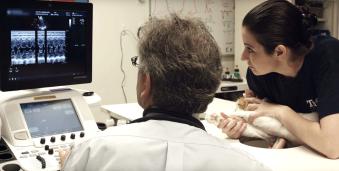
An echocardiogram being performed on a cat by Dr. John Rush. Echocardiography is an ultrasound of the heart. It is a noninvasive procedure, and your pet will lie awake on a padded table while a soft probe passes over their body. Some animals may need light sedation to calm them during the procedure. A patch of fur may be shaved to allow for a clearer picture of your pet’s heart.
An ultrasound forms a picture by sending harmless sound waves into the body and interpreting how they bounce back. An echo provides your veterinarian with clear views of your pet’s heart. The size of the heart chambers, the health of valves, the thickness and function of the heart walls, and other detailed anatomy is seen on the echo.
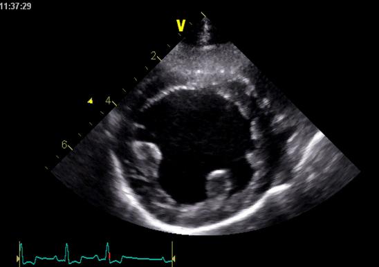
A picture from an echocardiogram (echo) of a dog heart An echo lets a cardiologist or ultrasound specialist view the heart pumping in real-time. The strength of the heart’s contraction is an important indicator of heart health. In heart disease, your pet’s heart may be contracting poorly, or the walls of the heart may be too thick.
Doppler technology lets your veterinarian track blood flow through the heart. Certain colors indicate specific directions of blood flow. Your veterinarian uses this to determine if there are any holes in your pet’s heart or any leaks at heart valves. Many heart diseases are due to leaky or stiff valves, and echocardiography enables observation of valve motion and function.
Echocardiography is a great tool for evaluating and diagnosing heart disease in pets, but it is just one tool. Your veterinarian may require other tests to guide treatment.
-
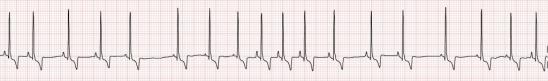
Electrocardiogram from a normal dog. An electrocardiogram (ECG) records the electrical activity of the heart. It is a safe, noninvasive procedure. Clips are attached over the legs and/or chest. Alcohol or another gel helps the clips connect to the skin so that the electricity generated by the heart can be recorded.
Your veterinarian may recommend an ECG if your pet has fainted (syncope) or has an irregular heartbeat. ECGs are also used in routine surgery or as part of some wellness exams.
Once attached, the clips start recording the heart’s electrical activity. Electricity washes through the heart in an organized fashion, so the heart contracts with each beat in an efficient and effective manner. The ECG might look like a bunch of squiggles, but these waveforms tell your veterinarian a lot about how the heart is activated and whether there is any concerning irregular heartbeat (cardiac arrhythmia).
Your veterinarian will begin analyzing the ECG by calculating the heart rate. Tachycardia describes a faster than normal heart rate. Two examples of fast heart rate that can be problematic are atrial fibrillation and ventricular tachycardia. These irregular heart rhythms can be associated with collapse or advanced heart disease, and specific treatment is usually required.
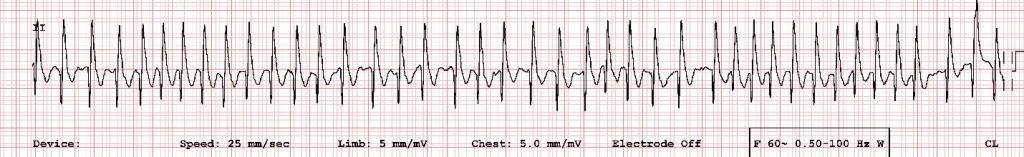
ECG from a dog with atrial fibrillation. 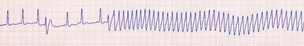
ECG from a dog with collapse due to ventricular tachycardia. If the heart rate is too slow, this is termed bradycardia. Very slow heart rates can be associated with collapse.
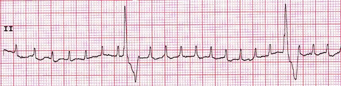
ECG tracing from a dog with fainting due to a slow heart rate (bradycardia). In some pets with slow heart rate and collapse, a pacemaker is needed to prevent sudden death.
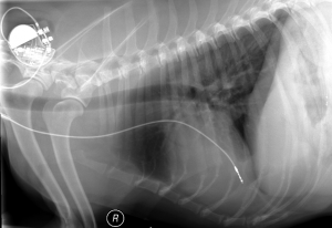
X-ray of a dog with a slow heart rate (bradycardia) that received a cardiac pacemaker to prevent collapse and sudden death. 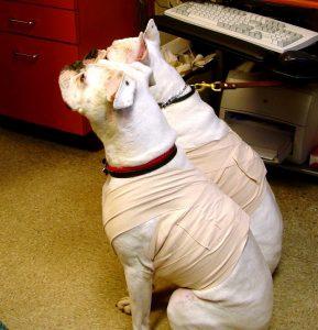
Two Boxer dogs wearing Holter monitors for 24-hour recording of the ECG. A specialized, portable ECG is a Holter monitor. This device records the electrical activity of your pet’s heart over 24-48 hours. Your pet wears the device like a harness. A Holter monitor is especially useful in a fainting animal. It can reveal whether an abnormal heart rhythm (arrhythmia) is causing the fainting. In animals with a known arrhythmia, the Holter monitor assesses the frequency and severity of the arrhythmia throughout the day.
*Note – ECG and EKG are both abbreviations for the same test. The difference in spelling is simply English (electrocardiography, abbreviated ECG) versus the German (elektrokardiographie, abbreviated EKG).
-
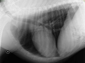
Chest x-ray from a normal dog. Note the heart size and the relatively black lung fields X-rays aren’t just for bones! They help your veterinarian assess the size, location and shape of your pet’s heart. Your veterinarian can also observe the lungs and blood vessels supplying these organs. X-rays (or radiographs) can help a veterinarian check for other causes of your pet’s symptoms. For example, your pet’s difficulty breathing may be due to a bone stuck in their throat, not heart disease.
To take an x-ray, your pet may or may not need to be lightly sedated. Sedation ensures your pet is calm and stationary during the x-ray. It also ensures the veterinary staff’s safety. If your pet moves during the x-ray, the images will be impossible to interpret. Your pet will be x-rayed lying down on their side and on their back or their stomach.
Your veterinarian will begin by analyzing the size, shape and location of your pet’s heart. In comparison to a healthy animal, an animal with heart disease may have an enlarged or unusually shaped heart on x-rays. Bulges in the heart shape can help pinpoint which heart chamber is enlarged. A markedly enlarged heart may indicate fluid buildup around the heart (pericardial effusion).
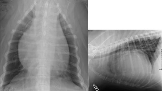
Two views of a chest x-ray from a dog with excess fluid around the heart, called pericardial effusion. One helpful tool to determine if the heart is enlarged is the vertebral heart score. This measurement compares the size of the heart to the size of the spine over the heart, which also shows up on a chest x-ray. An enlarged heart may help determine whether additional testing like an echo is needed, or whether certain drugs might be recommended.
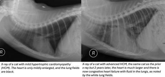
Your veterinarian will also evaluate the blood vessels connecting the heart and lungs. Your pet’s lungs can also be observed on x-ray. Heart failure in dogs and cats often leads to fluid buildup in or around the lungs. This fluid can look like a whiteness around or in the lungs. If the fluid in the lungs is due to heart disease then certain drugs, like diuretics, might be needed.
X-rays can tell your veterinarian a lot, but further testing is often still be necessary. A more complete diagnosis will help determine the best treatment for your pet.
-
Your veterinarian may recommend bloodwork to evaluate the heart and the other organ systems. Bloodwork includes a CBC (complete blood count) and blood chemistry panel. There is no single value on routine bloodwork specific for the heart, but the results help your veterinarian get a clearer picture of your pet’s overall health. Bloodwork lets your veterinarian evaluate whether heart failure or medications have caused any organ damage (especially the kidneys).
Your veterinarian may want to perform other blood tests, such as thyroid levels, taurine levels, BNP levels or troponin levels. Some of these tests examine specific substances related to heart function, sometimes called cardiac biomarkers. These substances are tested separately as they are not included on routine bloodwork.
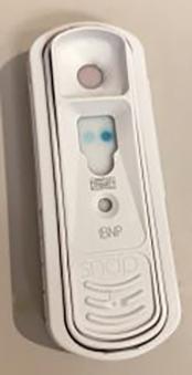
Bedside test for BNP from a cat with heart disease There are two main cardiac biomarkers, BNP and cardiac troponin I. A poorly functioning heart produces high levels of BNP (or NT-proBNP). BNP is a marker for heart stretch, dilation, or thickening. Most cats and dogs with moderate heart enlargement have some elevation of BNP, and the elevation is usually even more dramatic if they have heart failure. There is also a bedside test for BNP that can be done in cats.
A heart that was recently damaged, or one that is significantly diseased, will release cardiac troponin I into the blood. Cardiac troponin I can be measured in the blood, and elevated levels indicate damage to the heart.
If your pet is being treated for heart disease, your veterinarian may request periodic bloodwork to ensure medications are not damaging other organs. and troponin can be measured to help screen for heart disease. If your pet is genetically predisposed to heart disease, such as a Doberman pinscher or a Maine Coon Cat, these tests might help detect heart disease in its early stages. Elevation in these tests will usually trigger a recommendation for an echocardiogram (echo). Genetic testing for some heart diseases can be done, and one testing site is the North Carolina State University’s Veterinary Genetics Laboratory.
If your pet is being treated for heart disease, your veterinarian may request periodic bloodwork to ensure medications are not damaging other organs.
-
Elevation of blood pressure is sometimes called systemic hypertension. High blood pressure, or hypertension, is often caused by disease in the kidneys or other organs in dogs and cats. Measurement of blood pressure can be useful in animals suspected to have heart disease. High blood pressure causes the heart to work harder and can lead to heart enlargement. Treatment of high blood pressure with medications makes it easier for the heart to work efficiently.
On the other hand, some dogs and cats with heart disease can develop low blood pressure, called hypotension. Low blood pressure can cause weakness, lethargy, and loss of appetite. Hypotension can be due to advanced heart disease, although some of the medications used to treat heart disease can also create unwanted low blood pressure.
Your veterinarian may want to periodically check blood pressure in dogs or cats with heart disease. This is to make sure the heart is not worsening and that your pet has normal blood pressure. This information can help determine how heart medications might be adjusted.
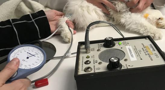
Blood pressure is measured with a small inflatable cuff, while a patient is laying on their side with gentle restraint. -
Cardiac catheterization is the insertion of a thin tube (a catheter) into a heart chamber or blood vessel. This procedure typically requires anesthesia for your pet’s safety. Although minimally invasive, it may be necessary for diagnosis and can be used for treatment of certain heart defects.
Through a small cut in the skin, your veterinarian will insert the catheter into a blood vessel. The catheter slides through the blood vessel into the heart. A specialized continuous x-ray (fluoroscopy) shows the catheter location. Your veterinary cardiologist may record the pressure in the heart chambers and blood vessels.
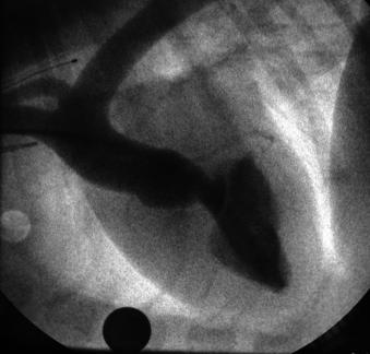
An angiocardiogram is like a chest x-ray that is obtained as dye is injected into the heart. The black pattern is from dye that appears in the left ventricle and the aorta. This dog has subaortic stenosis, a congenital defect that causes narrowing just below the aortic valve. Dye can be injected through the catheter to show the path of the blood through the heart. The dye outlines the heart chambers and blood vessels. This is called angiography or angiocardiography. It will help your veterinarian identify abnormal heart structures such as holes in the heart or abnormally narrow valves.

Cardiac catheterization procedures are performed sterilely, with pets asleep under general anesthesia. Some procedures to repair structural problems (closure of holes in the heart, balloon dilation of narrowed valves, and cardiac pacemaker implantation) use cardiac catheterization to access specific areas of the heart.