Heart Disease Basics
Heart Disease Resources: Basics | Symptoms | Diagnosis | Dogs | Cats | Treatments | Medications | Nutrition | Forms
A cat's or dog’s heart looks and acts much like your heart. It is an essential organ that performs important tasks. If you know how a healthy heart works, it will help you understand how it can fail to do its job. Understanding your pet’s unique heart condition can help you feel more empowered in your pet’s care.
If you suspect your pet may have a heart condition, please speak with your veterinarian or find a veterinary cardiologist.
-
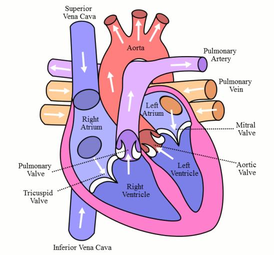
A cat or dog heart is a 4-chambered muscle that pumps blood through the body. The chambers of the heart are the right and left atrium and the right and left ventricle. Between the chambers are valves (flaps of tissues) that prevent the backflow of blood.
First, let’s talk about blood. The heart is responsible for pumping blood to the entire body. Blood contains high levels of oxygen and nutrients to feed the body. As blood flows, muscles and organs remove oxygen to use as fuel. After blood passes through the tissues, the low-oxygen blood returns to the right side of the heart. The right side of the heart pumps the blood through the lungs. In the lungs, the blood acquires fresh oxygen. The high-oxygen blood travels through the left side of the heart. The left side of the heart pumps blood out to the body. And the process repeats itself.
Next, we take a closer look at blood flow through the heart. Low-oxygen blood from the body enters the right atrium. The right atrium serves as a reservoir of blood, holding blood until a valve (called the tricuspid valve) opens. Blood flows across the open valve to the next chamber, the right ventricle. The right ventricle fills with blood and then contracts. The heart contraction pushes blood across the pulmonic valve and through the lungs. In the lungs, the blood picks up oxygen.
As blood returns from the lungs, the high-oxygen blood enters the left atrium. At the appropriate time, the mitral valve opens and blood flows into the left ventricle. The left ventricle is the most muscular chamber of the heart, and it serves as the main pumping chamber. When the left ventricle contracts it pushes blood across the aortic valve and into the aorta. The aorta disperses the high-oxygen blood to the entire body.
-
The body depends upon the heart to pump high-oxygen blood to organs, muscles, and tissue. All parts of the body need oxygen to function. When part of the heart is diseased, healthier parts of the heart may pick up the slack. This extra work can lead to thickening, thinning or enlargement of the heart muscle. Overall, disease in the heart causes the system to become inefficient.
Symptoms of heart disease may appear when the heart fails in its job as an efficient pump. One way the heart pump can fail is to be unsuccessful in pumping oxygenated blood to the body, leading to weakness or collapse. Another way the heart can fail is when fluid backs up behind the pump, causing fluid buildup in the lungs. Because the heart is so vital, symptoms can affect the entire body.
It is important to note that the main cause of heart disease in dogs and cats is usually very different from humans. Dogs and cats rarely develop coronary artery disease, also called atherosclerosis. This is a common human heart condition and is the main cause of heart attacks. Coronary artery disease in people typically causes the blood vessels that supply the heart with blood to become clogged or blocked.
Dogs and cats more often get problems with the heart muscle or heart valves, rather than the coronary arteries. Because of the different causes, heart disease in your pet can be different from what you might have seen or read about in people.
-
Many dogs and some cats with heart disease have a heart murmur. Murmurs are most often diagnosed while your veterinarian is listening to your pet’s heart. A normal cat or dog heart produces two sounds, which are sometimes referred to as “lub and dub”. The regular “lub dub” sounds happen as the heart is working normally and the valves are opening or closing.
Listen to normal dog heart sound here.
A murmur is an extra sound that is caused by abnormal or turbulent blood flow inside the heart.
Causes of heart murmur include almost any problem with the heart valves. The valves may be leaky, allowing blood to backflow, called regurgitation. This kind of is common in older small breed dogs that develop problems with their mitral valve.
Listen to heart murmur from a dog with mitral valve disease
Or the valves may be stiff with a narrow opening, called valve stenosis.
Listen to heart murmur from a dog with a narrowed aortic valve, called subaortic stenosis
Murmurs can also be associated with birth defects. One example of a defect is a hole in the heart or an abnormal connection of the blood vessels. This hole allows blood to flow between chambers that don’t connect in a normal heart.
If your pet has a murmur, your veterinarian will determine the intensity, location, and timing of the murmur. These findings can help pinpoint what part of the heart is diseased.
The intensity of a murmur is given a grade. Grade I/VI (or 1 out of 6 murmur) is the softest heart murmur and can be heard in some healthy animals. Grade VI/VI (or 6 out of 6 murmur) is the loudest murmur grade.
The grade, or loudness of the heart murmur, is sometimes an indication of the disease severity. Since the loudness, or grade, of the heart murmur does not always predict the severity of disease, additional testing is needed once a concerning heart murmur is noted by your veterinarian.
-
An abnormal heart rhythm, called an arrhythmia, is common in dogs and cats with heart disease. The heart pumps blood because of electrical impulses that course through the heart. Specific heart cells called pacemakers help direct this electricity. The sinoatrial (SA) node is the main pacemaker. It controls a regular heartbeat. A disruption of the pacemaker’s signal, or a spontaneous beat from someplace else in the heart, causes an arrhythmia.
An arrhythmia can be the heart beating too fast, called tachycardia. Or the heart can beat too slow, called bradycardia. Or the heart could have an irregular beat or pattern.
An irregular heartbeat reduces blood flow. This can cause collapse or fainting (syncope), weakness, and even death.
An electrocardiogram (ECG or EKG) analyzes the electrical activity of your pet’s heart. It will help your veterinarian diagnose your pet’s arrhythmia. ECG’s are non-invasive and safe.
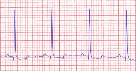
Electrocardiogram from a normal dog Arrhythmias are most often associated with heart disease. However, other conditions like pancreatitis or certain cancers can also cause arrhythmias.
-
While connected, heart disease and heart failure and congestive heart failure are distinct terms. They describe different levels of severity in heart malfunction.
Heart disease indicates part of the heart is faulty. For example, a valve might have a small leak due to a disease, but the leak is small enough that it does not yet cause any problems, and the heart still pumps blood normally. This animal has heart disease, but it is not showing any obvious symptoms and does not have heart failure yet.
In Heart failure, heart disease has progressed to the point where the heart is no longer doing a good job as a pump, and this is having a negative effect on the animal. The heart is failing in its job as a pump. If the heart fails to pump enough blood to the tissues, animals can develop weakness, limited ability to exercise, or even fainting.
Congestive heart failure is a special form of heart failure, where fluid backs up and starts to accumulate behind the heart. One analogy: If you live in a house with a basement that accumulates water, you might have a pump to keep the basement dry. If the pump is not working, fluid accumulates in the basement. In this analogy, the heart is the pump and the lungs are like the basement – as the heart fails, the lungs get filled with fluid, like a sponge filled with water (fluid in the lungs is also called pulmonary edema), which make it difficult for the animal to breathe. This can result in fast or difficulty breathing or a cough. This fluid accumulation in the lungs is usually best seen on a chest x-ray.
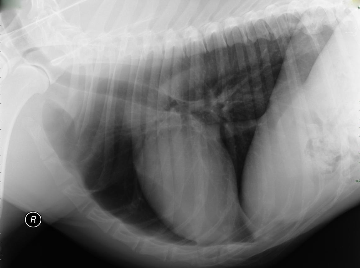
Chest x-ray from a normal dog. Note the heart size and the relatively black lung fields 
Chest x-ray from a dog with dilated cardiomyopathy and congestive heart failure. The heart is larger than normal and the whiteness in the lung fields is fluid in the lungs, called pulmonary edema. Some animals with congestive heart failure accumulate fluid in their belly (called ascites) or around their lungs (called pleural effusion).
An animal with heart disease is not always in heart failure, but an animal in heart failure always has heart disease. It is also important to note that fluid accumulation in the body is not always due to congestive heart failure.
HeartSmart Forms
- Heart Failure Monitoring Form
- Low-Sodium Diet for Dogs
- Low-Sodium Diet for Cats
- Treats and Nutritional Tips for Pets with Heart Disease
- FETCH Questionnaire: Assessing Quality of Life in Dogs with Heart Disease
- CATCH Questionnaire: Assessing Quality of Life in Cats with Heart Disease
- FETCH and CATCH Questionnaire Use Guidelines