Field Diagnostic Imaging
-
Tufts Veterinary Field Service performs equine radiography (X-rays) on-farm and by haul-in appointments in order to provide owners with immediate results regarding pain and lameness in teeth, neck, back and hooves.
With many lameness situations, veterinarians take radiographs as a first line method of diagnosis. Most commonly, the veterinarian looks for evidence of a joint being inflamed for long enough whereby the bones will start to change, either by dissolving away or laying down more (bone spurs), sometimes a mix of both. These are called destructive or productive lesions but are more commonly known as osteoarthritis. Other problems that X-rays can uncover include osteochondritis (a defect in the cartilage overlaying the bone), injuries, developmental abnormalities or congenital deformities and bone disease.
The veterinarians at Tufts Veterinary Field Service also work closely with farriers as they trim and balance the horse's hooves, offering on-the-spot guidance and consultations for horses with laminitis and corrective shoeing needs.
If calling to schedule an X-ray appointment, please identify that service when calling, as there is one available machine in the practice.
-
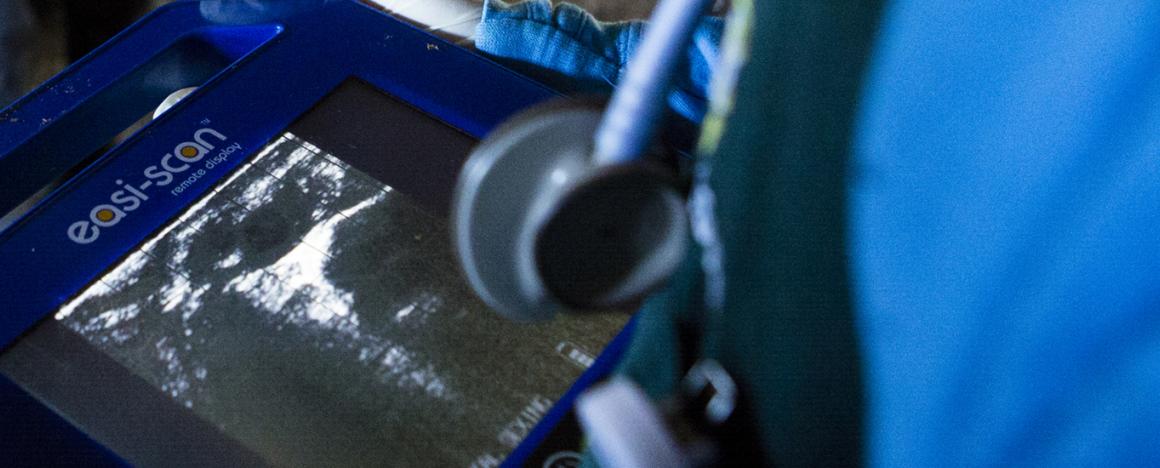
Many horse owners are familiar with the usage of diagnostic ultrasound to confirm the pregnancy of a horse, however, ultrasound it is also employed by Tufts Veterinary Field Service to help evaluate injuries and inflammation in tendons, ligaments and other soft tissues, including tissues around joints.
A handheld probe emits high-frequency sound waves that travel into the horse's body. Ligaments, tendons, bone and other tissues reflect the sound waves in different degrees, based on their density. The scan is non-invasive when capturing the echoes and creates a detailed image of the different soft tissue structures.
Ultrasound can also be performed in abdominal and thoracic areas, as well as to help veterinarians obtain crucial information about a horse’s eye when a routine ophthalmologic exam is not possible due to conditions such as severe eyelid swelling.
If calling to schedule an ultrasound appointment, please identify that service when calling, as there is one available machine in the practice. Ultrasounds may done either on-farm or as a haul-in service at the clinic in Woodstock, CT.
-
When problems with coughing or breathing arise, or when a horse is losing weight or not eating well, endoscopy is a useful tool. Upper and lower endoscopy involve examining the airways (trachea) and pharyngeal regions, including the guttural pouches. Gastroscopy involves passing the endoscope down the esophagus to the stomach.
Using a three-meter scope, the veterinarian most commonly evaluates the horse for gastric ulceration suspicions. With the ability to acquire an image right in the field to interpret and diagnose, endoscopy also allows the veterinarian to detect abnormalities such as infection, cancerous growths or foreign bodies.
Tufts Veterinary Field Service is capable of performing equine endoscopies either on-farm or as a haul-in service to their clinic in Woodstock, CT.
Because Tufts Veterinary Field Service enjoys a close working relationship with Cummings School of Veterinary Medicine at Tufts University in Grafton, MA, the veterinarians will reach out to their esteemed colleagues at Cummings School should questions arise regarding the outcome of an endoscopy.
A horse must be fasted the night before a gastroscopy (stomach endoscopy) appointment.