Seeing Further
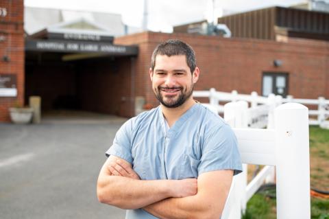
As a veterinary technician working in nuclear medicine and CT, Josh Peters has the unique capability to help reveal health issues in animals of all sizes
When horses experience a decrease in their athletic performance, it could indicate an injury or an underlying disease. Imaging plays a central role in accurately and rapidly diagnosing the problem.
Our Imaging Service is one of the select few in the country offering the full-range of imaging services with advanced technology and a team of board-certified radiologists and dedicated equine ultrasongraphers.
Our board-certified clinicians work in collaboration with our radiologists to interpret images in real time for a swift and accurate diagnosis enabling them to solve both common and puzzling cases. Tufts Equine Center is fully equipped with digital radiography, fluoroscopy, a dedicated equine ultrasound suite, nuclear medicine, a Helical Multislice CT unit and the only 1.5 high-strength Tesla Magnet Resonance Imaging (MRI) unit in New England. In addition to offering imaging services within the hospital, we also provide consultations to referring veterinarians who are seeking a second option on their own medical imaging studies.
All of our images are acquired and stored digitally and can be transferred easily to referring veterinarians and horse owners in digital or printed format. We are always available to discuss with horse owners and veterinarians the type of imaging scan that will work best for their individual needs.
Learn more about the advanced imaging services we provide:
CT images can lead to a quicker diagnosis and more detailed analysis of lameness and poor performance. Tufts Equine Center is one of the few places with the ability CT horses owing to our specially designed and constructed table that allows us to position horses to image their heads and legs. Our team of board-certified clinicians, radiologists and anesthesiologists work in collaboration to utilize this powerful and versatile diagnostic tool.
CT offers ideal imaging of the equine head to detect dental, nasal and sinus disease as well as fracture depiction. It is also preferred for identifying tendon and ligament injuries and bone infections in foals. Because of its rapid acquisition time, it is also beneficial in select cases for fracture assessment prior to undertaking repair, as well as in pre-operative planning to have more insight in advance of the surgical procedure.
Veterinarians at the Tufts Equine Center can identify causes of lameness, neurological conditions and other illnesses that affect horses by using the first and only equine-capable, high-strength (1.5 Tesla) magnetic resonance imaging (MRI) technology in New England. The MRI is similar to that which is used on human patients. Many veterinarians agree that MRI should be part of a full diagnostic lameness workup, used in conjunction with findings from the horse’s primary veterinarian.
Our equipment is instrumental in diagnosing a variety of equine musculoskeletal conditions, many of which cannot be detected using other forms of diagnostic imaging. Horses are anesthetized and placed on a non-magnetic table that has been specially designed and built for our equine patients. The MRI uses strong magnetic fields and radio frequency pulses to produce images with superior bone and soft tissue detail. It is used to diagnose problems in many different structures in the horse, including soft tissue lesions that cause lameness, brain lesions that may cause seizures, and small tendon tears within the horse's hoof. Clinicians, radiologists, anesthesiologists and other staff members work as a team to obtain MRI images and determine a diagnosis. Our high-strength MRI capabilities and the interpretive power of our equine veterinary team are unique in New England.
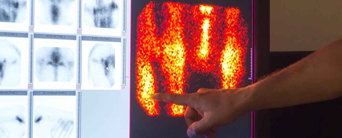
Nuclear scintigraphy, also known as bone scanning, is a form of diagnostic imaging designed to detect areas of increased bone metabolism that may signal orthopedic diseases. It is especially helpful in identifying multiple areas in the skeletal system that may be contributing to lameness. Bone scans enable our veterinarians here at Tufts Equine Center to look at regions of the body otherwise inaccessible by other imaging modalities, such as the back, pelvis and hips of the horse.
During the procedure, the patient is injected with a radioactive substance (radioisotope) that accumulates in bones and soft tissue. After the injection, the horse is placed in a stall for a few hours to allow the isotope to settle into the affected areas. The horse is then scanned with a gamma camera. The gamma camera detects the isotope signal as it emanates from the horse and produces an image that highlights specific areas of inflammation. From these images, the clinician can determine the affected areas and narrow down the potential causes of lameness. Horses are kept in the hospital for 24 hours until the radioactivity has cleared from their bodies.
Our equine surgery suites are equipped with a 9800 OEM General Electric C-Arm. The technology allows equine surgeons to accurately place orthopedic implants during the surgical procedure. The unit is designed to perform contrast vascular studies similar to those performed on human patients.
Digital radiography is another form of X-ray imaging commonly used for diagnosing problems in patients. In the large animal routine radiographic suite, we take images of bone and soft tissue using digital sensors as opposed to the traditional photographic method. By using this digital format, we can manipulate the images on a computer, magnify lesions and enhance specific areas to better visualize the problem. The digital method also allows us to easily share the images electronically with colleagues and clients, as well as the clients' primary veterinarians.
While portable radiographic equipment is often limited to only the legs of a horse, our high-powered unit allows our clinicians and staff at Tufts Equine Center to take radiographs of a horse’s chest, neck, back and head.
We also rely on myelography, a radiographic examination that uses injected contrast material to detect diseases of the spinal cord. A myelogram is performed on an anesthetized horse to detect compressive lesions of the cervical vertebra through flexion of the head and neck.